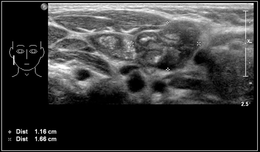
Ultrasound Anatomy Of The Neck
Related posts of neck muscle anatomy ultrasound knee muscle anatomy pictures. A common neck ultrasound is ultrasound of the thyroid which uses sound waves to produce pictures of the thyroid gland within the neck.

The purpose of this study was to assess the patency of the vessels the degree of tortuosity velocity of blood flow at different levels of vessels detection of atherosclerotic plaques and thrombi that threaten the development of stroke.

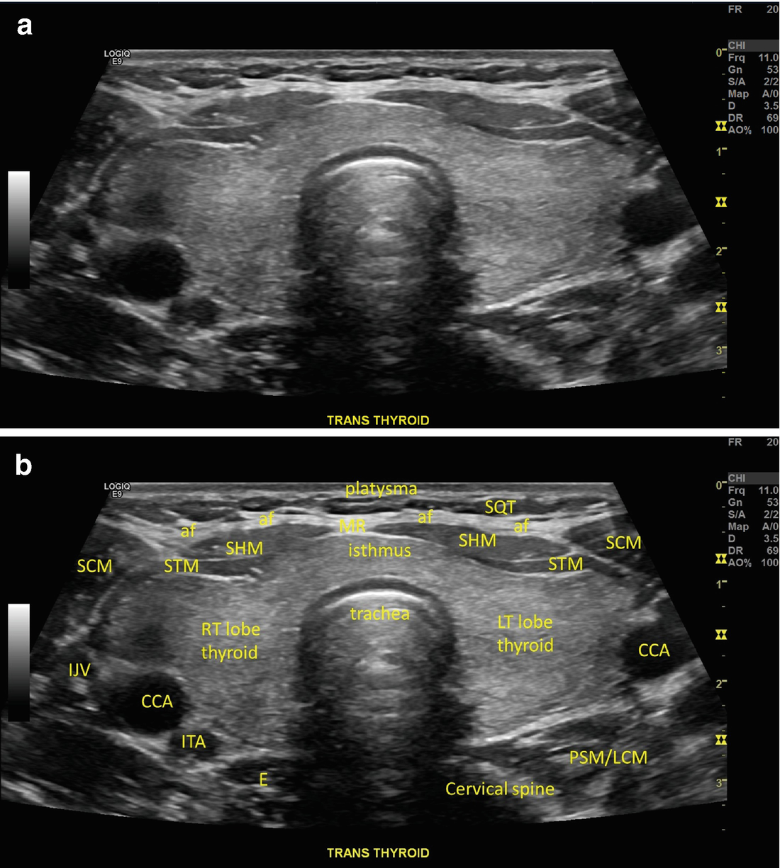
Ultrasound anatomy of the neck. Knee muscle anatomy pictures 12 photos of the knee muscle anatomy pictures knee muscle anatomy images knee muscle anatomy pictures human muscles knee muscle anatomy images knee muscle anatomy pictures. Diagnostic accuracy in suitably trained hands. It surrounds the front and sides of the visceral space and is related posteriorly to the carotid space.
Find out more from alaska family sonograms. While the vast majority of patients are supine on the exam table with a pillow supporting the shoulders to allow gentle neck extension keep in mind that some patients have beautiful anatomy d that allows ultrasound exam even in a sitting position. The most common and most popular type of ultrasound of the neck is an ultrasound of the neck vessels also called duplex receptacles duplex scanning of vessels doppler ultrasound.
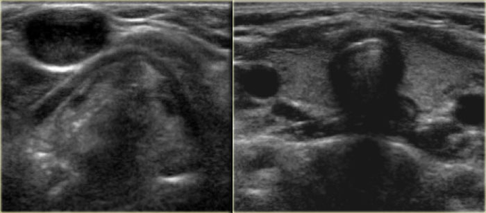
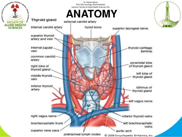
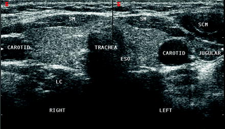
A neck ultrasound is performed to diagnose potential problems of the thyroid lymph nodes and carotid arteries. This article reviews the ultrasound features of the structures located in the infrahyoid region of the neck. For the head or neck evaluation a high resolution small part transducer with higher frequencies is generally used.
Optimal positioning and exposure of the neck for ultrasound of the thyroid and parathyroid glands a b and lateral neck for lymph node examination and mapping c. It does not use ionizing radiation. The anterior cervical space is located deep to the strap muscles and sternocleidomastoid muscle.
 The Radiology Assistant Infrahyoid Neck
 Normal Neck Anatomy And Method Of Performing Ultrasound
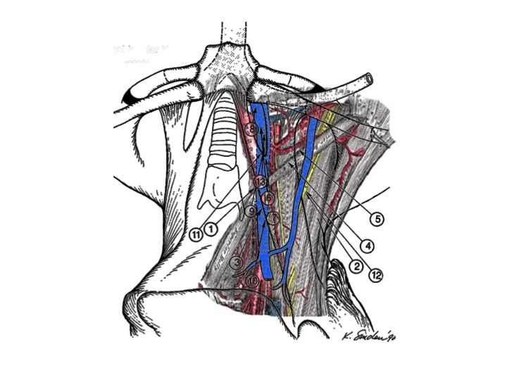
 Chapter 26 Triangles And Root Of The Neck The Big Picture
 Normal Neck Anatomy And Method Of Performing Ultrasound
 Figure 3 From Sonographic Anatomy Of The Neck The
 Chapter 25 Overview Of The Neck The Big Picture Gross
 Objectives Cervical A
 Neck Anatomy Ultrasound Sonography Thyroid Ultrasound
 Pancreas Normal Anatomy Virginia S Sonography Site
 Thyroid Ultrasound
 What Is An Anatomy Ultrasound During Pregnancy Babymed Com
.jpg) Ent Ultrasound Applications
Ent Ultrasound Applications
 Normal Neck Anatomy And Method Of Performing Ultrasound
 Diagnostic Ultrasound Head And Neck 9781937242169
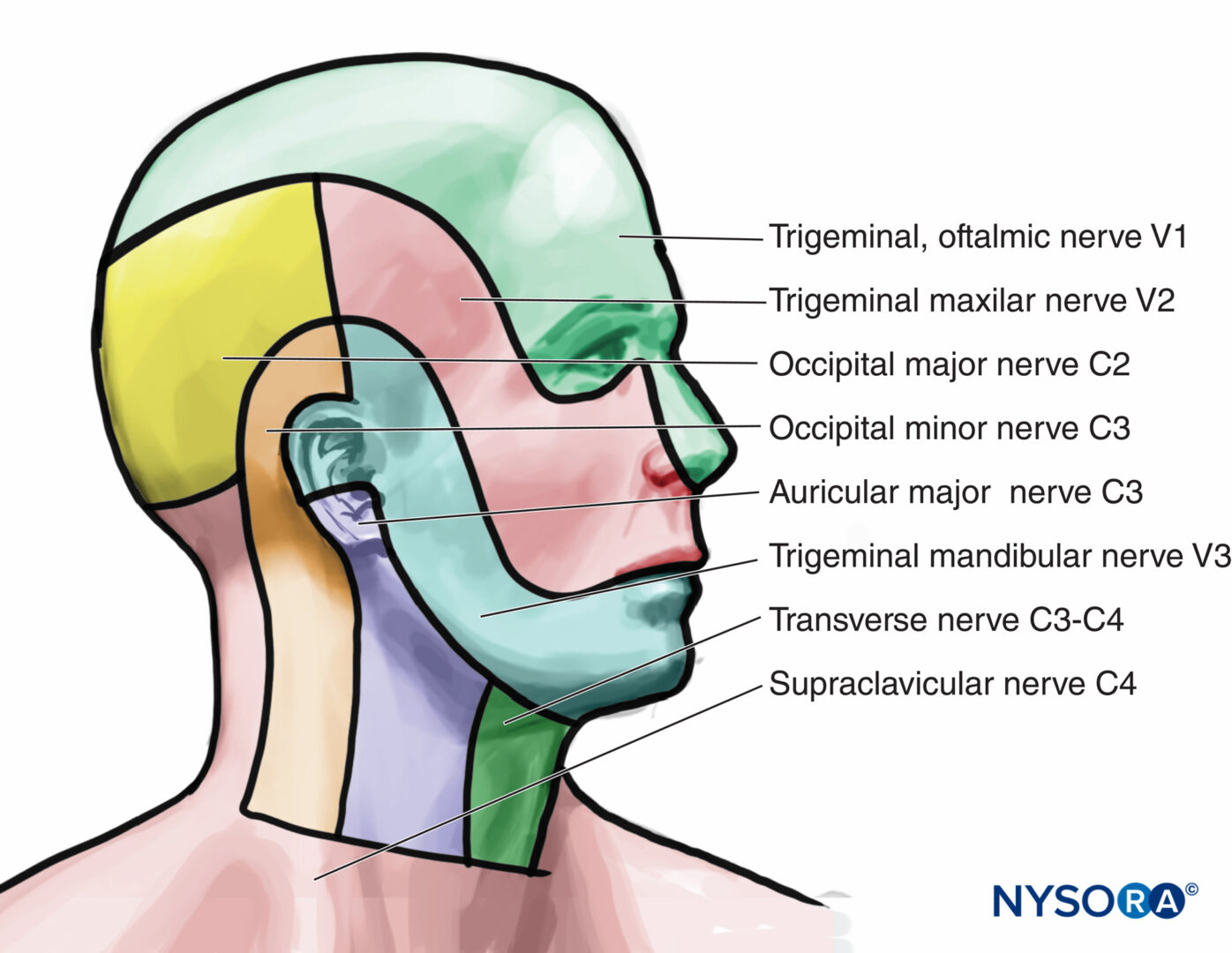 Ultrasound Guided Cervical Plexus Block Nysora
 The Radiology Assistant Infrahyoid Neck
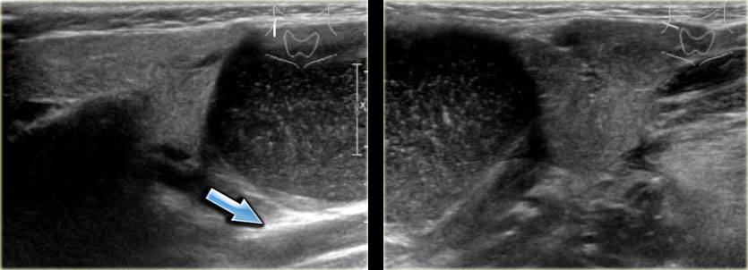
 Post Operative Ultrasound Evaluation Of The Neck In Thyroid
 Practical Anatomy Of Head Neck 4
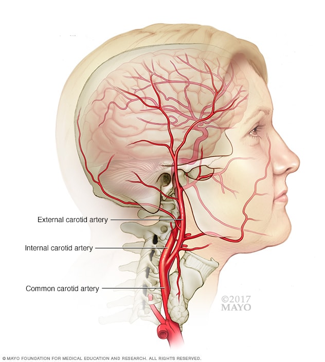 Carotid Ultrasound Mayo Clinic
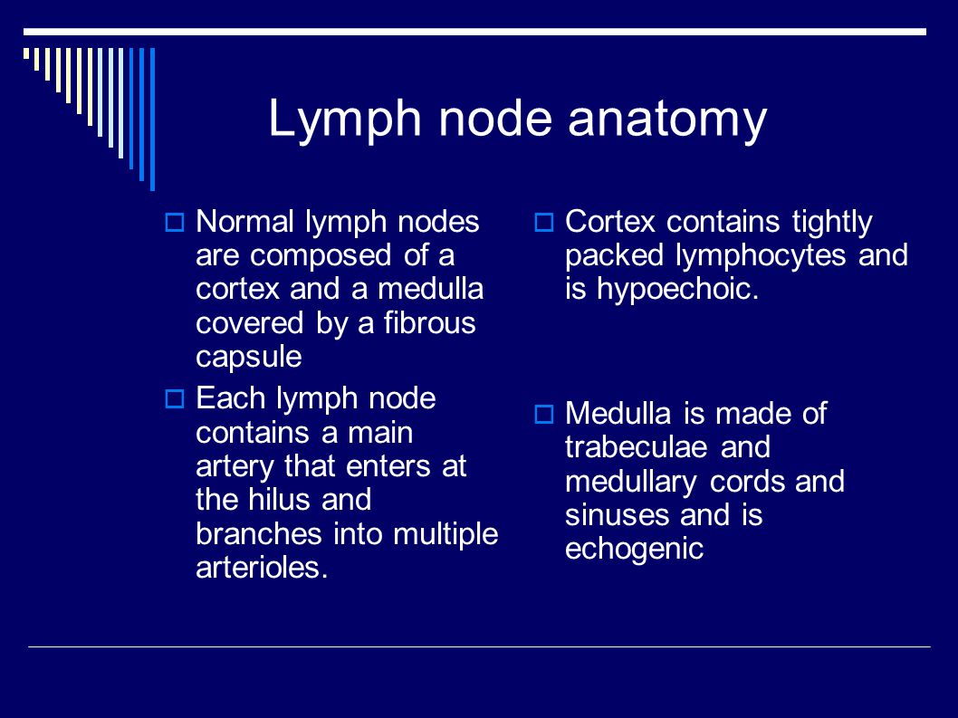
Normal Cervical Lymph Node Appearance And Anatomic Landmarks
 Chapter 4 Ultrasound Of The Neck Thyroid And Parathyroid
 Dr Matt Bull On Twitter Case Neck Ultrasound Anatomy
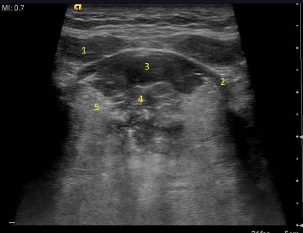
 Ultrasound Anatomy Of The Neck The Infrahyoid Region
 Ultrasound Anatomy Of The Neck The Infrahyoid Region

 The Radiology Assistant Neck Masses In Children
 Ultrasound Of The Shoulder
 Figure 9 From Sonographic Anatomy Of The Neck The
Belum ada Komentar untuk "Ultrasound Anatomy Of The Neck"
Posting Komentar