Womb Anatomy
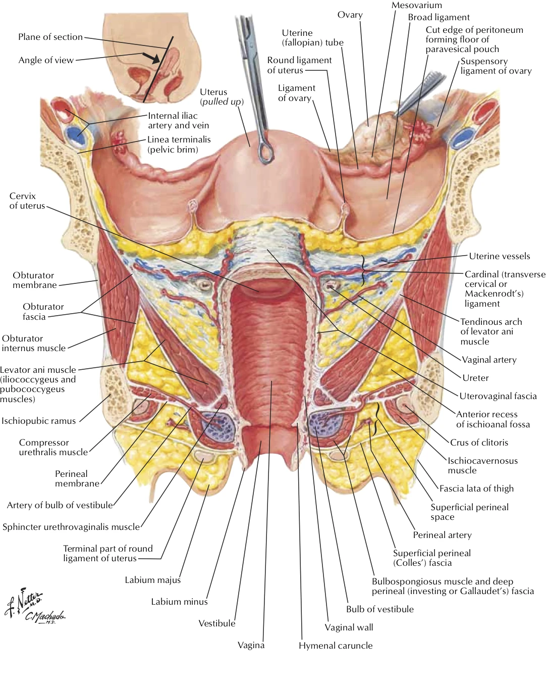
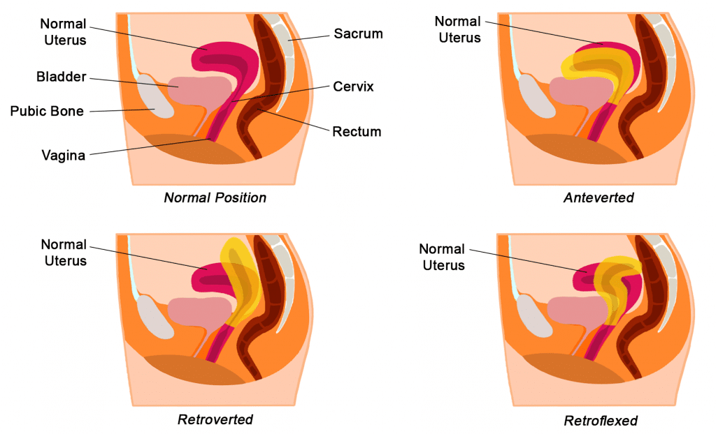
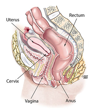
Uterus also called womb an inverted pear shaped muscular organ of the female reproductive system located between the bladder and rectum. The cervix is a cylinder shaped neck of tissue that connects the vagina and uterus.
The uterus performs multiple functions and plays a major role in fertility and childbearing.

Womb anatomy. The uterus or womb is shaped like an inverted pear. It functions to nourish and house the fertilized egg until the unborn child or offspring is ready to be delivered. Womb is an enlarged organ.
The uterus has di. In the human embryo the uterus develops from the paramesonephric ducts which fuse into the single organ known as a simplex uterus. This organ is able to change in shape as muscles tighten and relax to make it possible to carry a fetus.
It is located posterior to the urinary bladder and is connected via the cervix to the vagina on its inferior border and the fallopian tubes along its superior end. The middle layer or myometrium makes up most of the uterine volume and is the muscular layer. Located at the lowermost portion of the uterus the cervix is composed primarily of fibromuscular tissue.
Difference between womb and uterus definition. The uterus also known as the womb is a hollow muscular pear shaped organ found in the pelvic region of the abdominopelvic cavity. The uterus or womb is a major female hormone responsive secondary sex organ of the reproductive system in humans and most other mammals.
The anatomy of the uterus consists of the following 3 tissue layers see the following image. It is connected distally to the vagina and laterally to the uterine tubes. The uterus is a thick walled muscular organ capable of expansion to accommodate a growing fetus.
The uterus also known as the womb is the hollow organ in the female reproductive system that holds a fetus during pregnancy. The uterus otherwise known as the womb is the female sex organ that carries a huge significance in many species survival ours included. Uterus is smaller than the womb.
The uterus itself is a hollow organ that is shaped in the form of a pear and interestingly enough measures about that size. Womb is responsible for the development of the embryo. In the human the lower end of the uterus the cervix opens into the vagina while the upper end the fundus is connected to the fallopian tubes.
It is neatly tucked into the pelvic area of most mammals and of course in humans. The inner layer called the endometrium is the most active layer and responds to cyclic ovarian. Womb refers to an organ of a female mammal where the offspring is conceived.
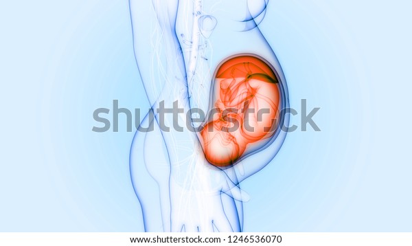
It is connected distally to the vagina and laterally to the uterine tubes. It is within the uterus that the fetus develops during gestation.
 Fetus Baby In Womb Anatomy Royalty Free Stock Image
 Vintage Anatomy Of A Human Infant In Womb Coffee Mug
 Female Reproductive System Everyday Health
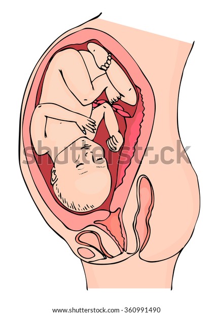 Ppregnant Woman Fetus Womb Anatomy Fetus Stock Image
 Uterus And Ovaries Preview Human Anatomy Kenhub
 Pregnant Womb Fetus Anatomy Cookie Cutter
 The Uterus Anatomy Of The Uterus Physiology Of The
 Fetus In Womb Medical Model With A Cross Section Of The Inner Organ With Red And Blue Arteries And Adrenal Gland As A Health Care And Medical Of The
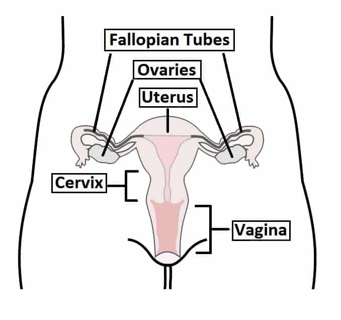 The Uterus Structure Location Vasculature Teachmeanatomy
 Cervix Definition Function Location Diagram Facts
 The Female Reproductive System Boundless Anatomy And
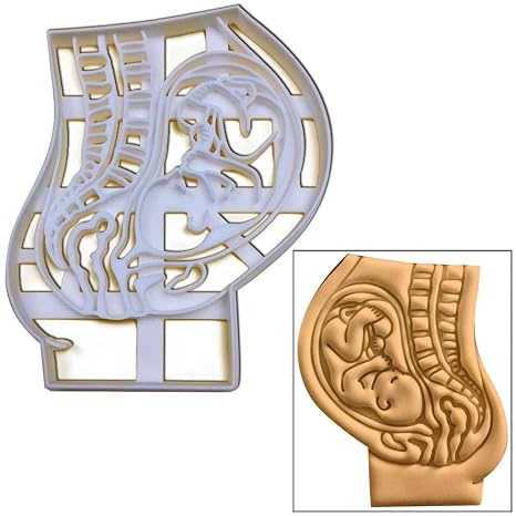 Amazon Com Pregnant Womb With Fetus Cookie Cutter 1 Pc
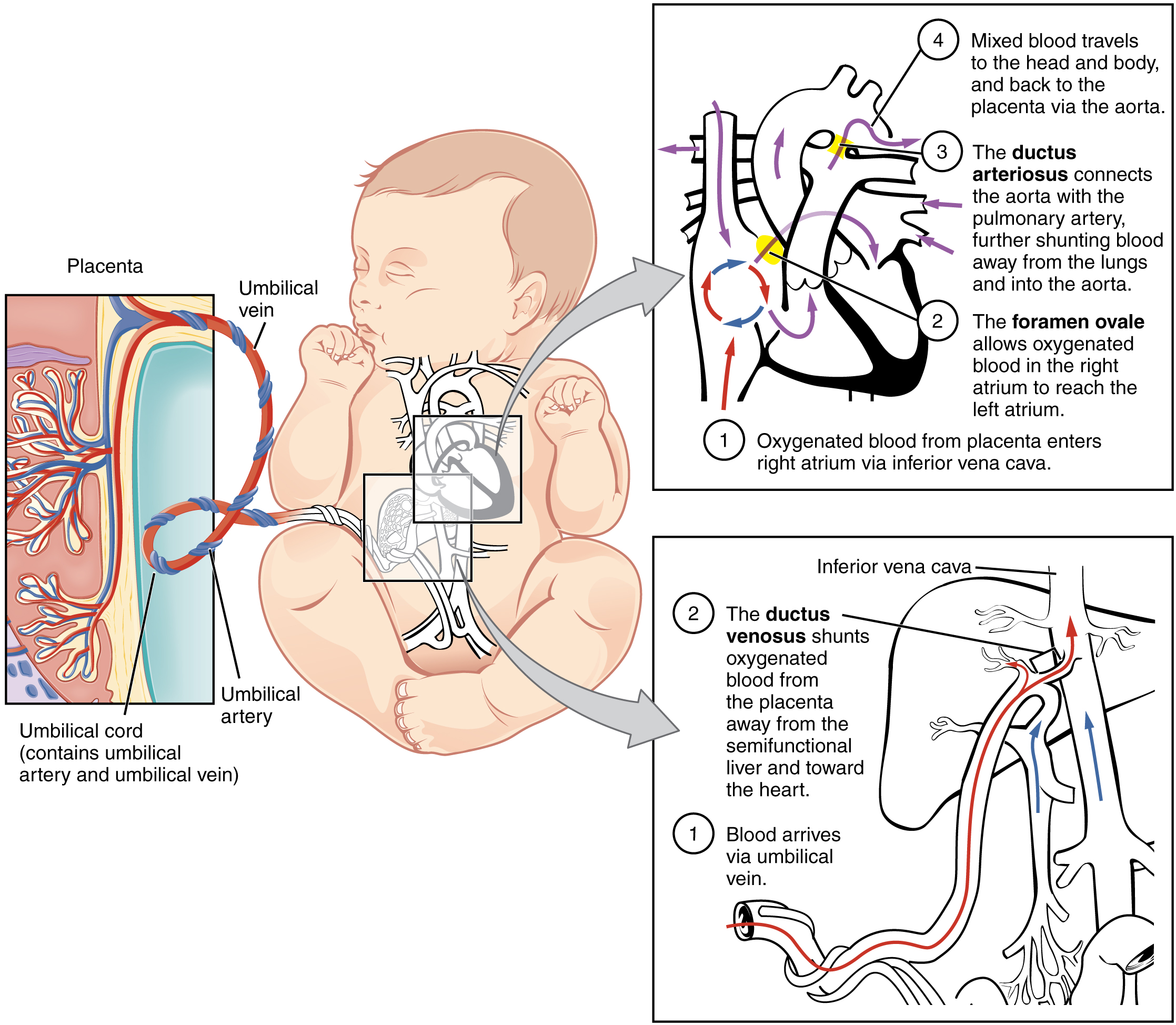 28 3 Fetal Development Anatomy And Physiology
 Viscera Uterus Ranzcrpart1 Wiki Fandom
 The Uterus Structure Location Vasculature Teachmeanatomy


 Fetus Baby In Womb Anatomy Buy This Stock Illustration
 What Organs Are Near The Uterus Quora
 Fetus Baby In Womb Anatomy Stock Illustration Illustration
 Fetus Baby Womb Anatomy 3d Stock Illustration 1246536070

Belum ada Komentar untuk "Womb Anatomy"
Posting Komentar