Anatomy Of The Ciliary Body
A part of the uvea. The ciliary body is part of the uvea the layer of tissue that delivers oxygen and nutrients to the eye tissues.
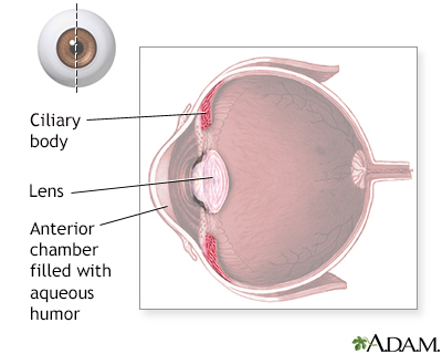
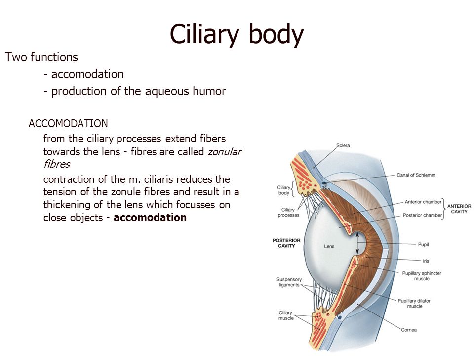
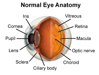
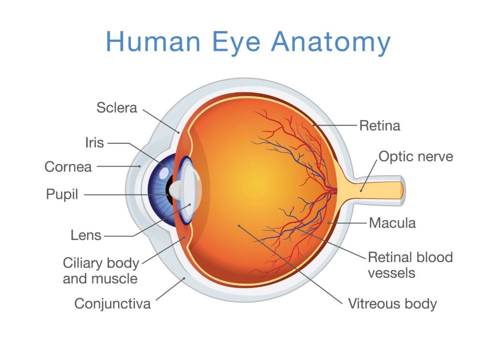
The ciliary body is a part of the eye that includes the ciliary muscle which controls the shape of the lens and the ciliary epithelium which produces the aqueous humor.
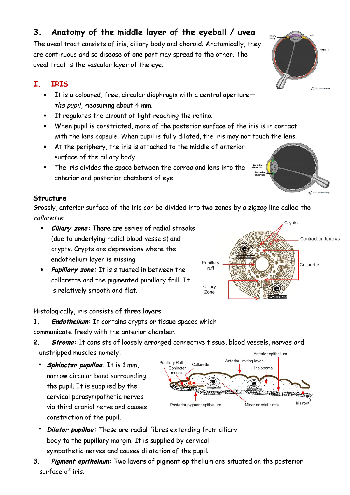
Anatomy of the ciliary body. This process is called accommodation. The ciliary body is the site of aqueous humor production. It also contains the ciliary muscle which changes the shape of the lens when your eyes focus on a near object.
Ciliary body laser therapy in ophthalmology. Anatomy of ciliary body. The vitreous humor is produced in the non pigmented portion of the ciliary body.
Anatomy of ciliary body ciliary processes anterior chamber angle and collector vessels 1. The ciliary body is the tissue which covers the inner part. Skip navigation sign in.
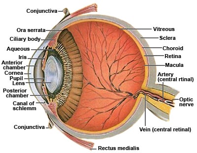
The ciliary body is the forward continuation of the choroid. The iris inserts into the anterior side of the ciliary. Ciliary body histology answers.
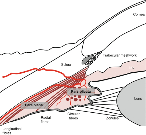
On cross section the ciliary body has the shape of a right triangle approximately 6 mm in length where its apex is contiguous with the choroid and the base close to the iris. Safety evaluation of ocular drugs. The ciliary body faces the anterior chamber posterior chamber and vitreous cavity and is lined by two neuroepithelial layers a non pigmented layer internally and a pigmented layer externally.
Ultrastructure of the ciliary processes. The outer layer of epithelium is pigmented but the inner layer which is in contact with the aqueous is non pigmented. The ciliary body produces aqueous humor.
It is a muscular ring triangular in horizontal section beginning at the region called the ora serrata and ending in front as the root of the iris. Separates epithelium from ciliary muscle thickest in pars plicate anteriorly joins ct or iris posteriorly joins stroma of choroid and collagenous and elastic layer of bruchs passage for bvs and nerves fenestrated bvs. The ciliary body is a part of the eye that includes the ciliary muscle which controls the shape of the lens and the ciliary epithe.
The ciliary body in cross section is cylindrical in rodents. The ciliary body joins the ora serrata of the choroid to the root of the iris. The ciliary body is a circular structure that is an extension of the iris the colored part of the eye.
The ciliary body produces the fluid in the eye called aqueous humor.
 External Anatomy Of The Eye Ppt Video Online Download
 Ocular Anatomy The Ciliary Body Ciliary Muscle Muscle
Anatomy Of The Eye Richmond Eye Associates
 The Eyes Canadian Cancer Society
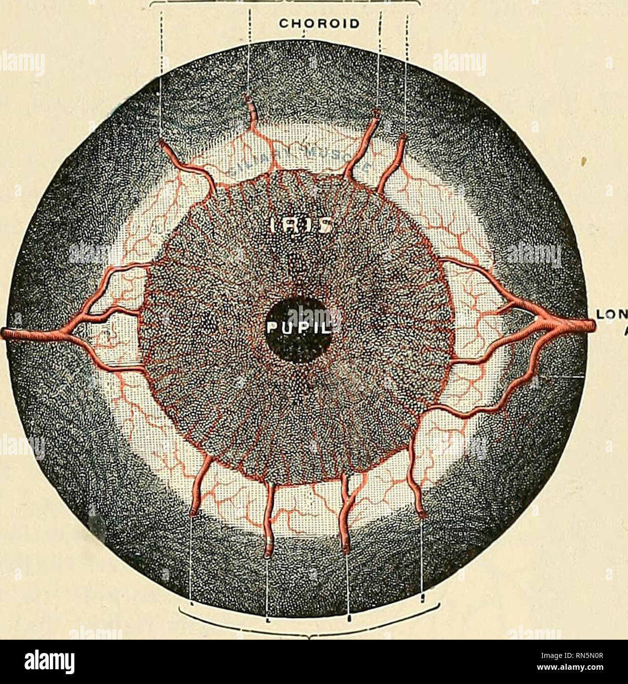 Anatomy Descriptive And Applied Anatomy The Choroid
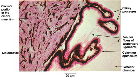 Anatomy Atlases Atlas Of Microscopic Anatomy Section 1 Cells
 Anatomy Of The Ciliary Body And Outflow Pathways Ento Key
 Eye Anatomy How Youniggasook Od Ciliary Body Retina Iris
 3 Anatomy Of The Middle Layer Of The Eyeball Uvea Auto
 Ciliary Muscle An Overview Sciencedirect Topics
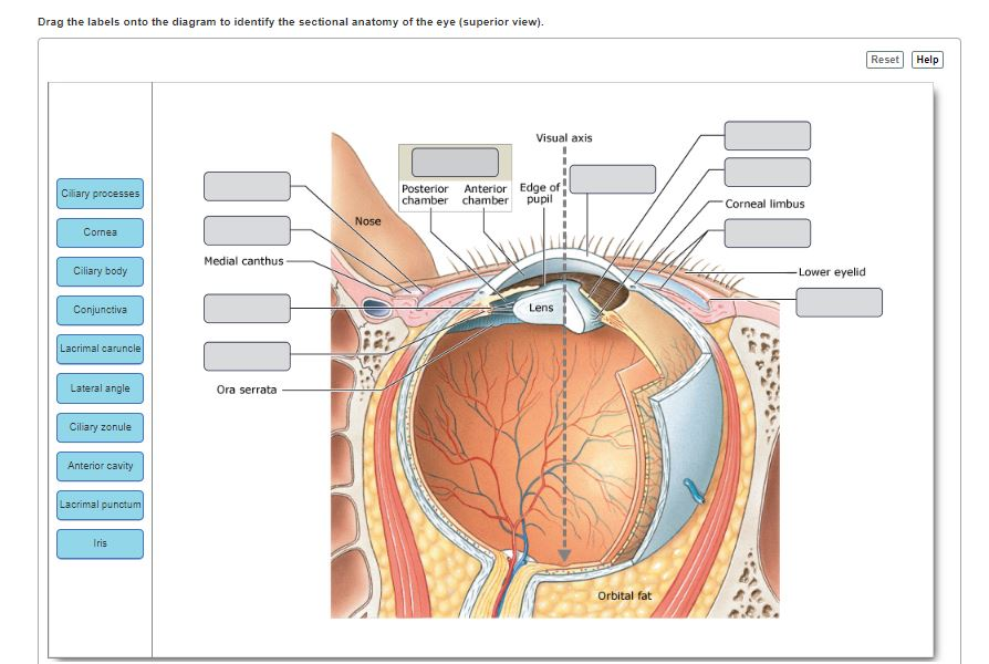 Solved Drag The Labels Onto The Diagram To Identify The S
 Eye Anatomy Glaucoma Research Foundation
 The Ciliary Body And Aqueous Fluid Formation And Drainage
 Anatomy Of The Ciliary Body And Outflow Pathways Ento Key
 Instant Anatomy Head And Neck Areas Organs Eye Orbit
 Anatomy Of Ciliary Body Ciliary Processes Anterior Chamber
Anatomy Of Uveal Tract By Dr Parthopratim Dutta Majumder
 Orbits And Eyes Anatomical Illustrations
 Vision And The Eye S Anatomy Healthengine Blog
 Aqueous Humor Flow And Function Diagram And Glossary
 Ciliary Body Information Mount Sinai New York
 Chapter 2 Anatomy Of The Eye And Common Diseases Affecting
 Lens And Ciliary Body Anatomy Histology And Action Preview Kenhub
 Anatomy Of The Eye Red Rover Ventures
 Eye Anatomy Ocular Anatomy Vision Conditions Problems
 Anatomy And Structure Of The Eye Brightfocus Foundation
 Eye Iris Pupil Ciliary Body Ppt Video Online Download
 Anatomy Of The Eye Children S Wisconsin



Belum ada Komentar untuk "Anatomy Of The Ciliary Body"
Posting Komentar