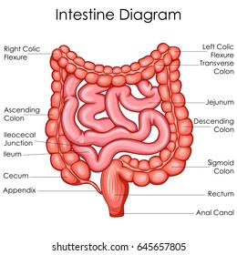
Cecum Anatomy
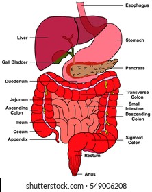
The cecum is the most proximal part of the large intestine and is located between the ileum distal small bowel and the ascending colon. The word cecum stems from the latin caecus meaning blind.
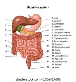
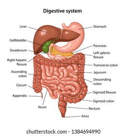
 Digestive System Structure Of The Digestive System
It is typically located on the right side of the body.

Cecum anatomy. Having served as a site for cellulose digestion in our ancestors the cecum now simply acts as a reservoir for chyme which it receives from the ileum. While the cecum is usually intraperitonea. The cecum the first part of the large intestine is a sac with a closed end that occupies the right iliac fossa the hollow of the inner side of the ilium the upper part of the hipbone.
The mucosal layer forms the innermost layer of the cecum comprising of mucous membrane containing various goblet cells. The goblet cells are responsible for releasing mucus which helps in lubricating the cecum walls. It is also separated from the colon by the cecocolic junction.
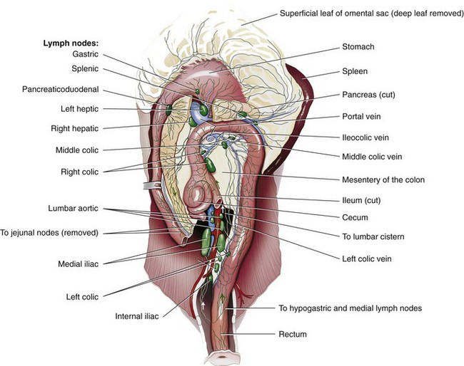
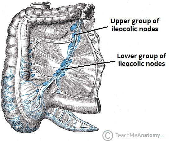
Muscularis strongly pronounced inner circular musculature outer longitudinal musculature almost. Lymph from both the appendix and cecum drain into the ileocolic lymph nodes. Mucosa columnar epithelium with crypts contains goblet and endocrine cells.
Submucosa with blood vessels and lymph nodes. The cecum is supplied by the ileocolic artery which is a terminal branch of the superior mesenteric artery. It receives chyme from the ileum and connects to the ascending colon of the large intestine.
The cecum comprises of four different layers of tissue all having distinct functions. The microscopic structure of the cecum is equal to that of the colon. Histology and anatomy of the cecum.
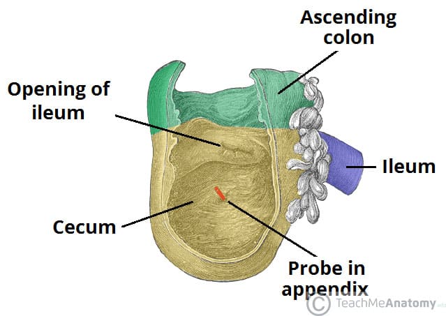
It is separated from the ileum by the ileocecal valve or bauhins valve. Guarding the opening of the ileum the terminal portion. The cecum or caecum is a pouch within the peritoneum that is considered to be the beginning of the large intestine.
The ileocolic artery gives rise to the appendicular artery to supply the appendix.
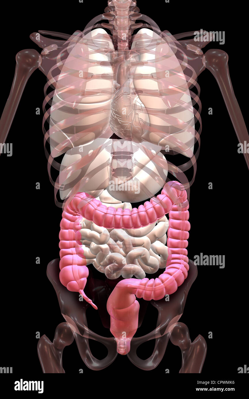
 The Large Intestine Human Anatomy
 Colon Veterian Key
 The Vermiform Appendix Is The Inferior Extension Of The
 The Cecum Position Vasculature Teachmeanatomy
 Details About Digestive System Stomach Anatomy Model Large Intestine Cecum Teaching Supplies
 The Appendix Retrocecal Arterial Supply Appendicitis
 Royalty Free Cecum Stock Images Photos Vectors Shutterstock
 Appendicitis Vector Illustration Labeled Appendix Inflammation
 Anatomy Of The Large Intestine
 The Cecum Position Vasculature Teachmeanatomy
 Royalty Free Cecum Stock Images Photos Vectors Shutterstock
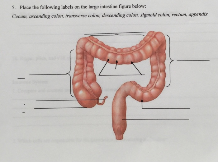 Solved 5 Place The Following Labels On The Large Intesti
 Foramen Cecum Of Tongue Anatomy Significance Abnormality
 Large Intestine Anatomy And Digestion Nutrition Biology
 Human Digestive System Stomach Anatomy Model The Large
 Anatomical Illustration Showing The Appendix Cecum And
 Large Intestine Course Hero
 Large Intestine Structures 1 Cecum 2 Ileocecal Valve 3
 Cecum Wikipedia
 Lower Gastrointestinal Tract Sciencedirect
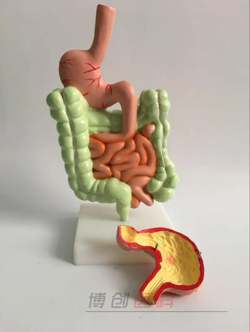 Details About Human Digestive System Stomach Anatomy Model The Large Intestine Cecum Rectum
 The Colon Ascending Transverse Descending Sigmoid
 Definition Of Cecum Nci Dictionary Of Cancer Terms
 Bilder Stockfotos Und Vektorgrafiken Blinddarm Shutterstock
 Anatomy 1 C2 L9 Large Intestine
 Royalty Free Cecum Stock Images Photos Vectors Shutterstock
 Midgut Distal Half Of The Duodenum Jejunum Ileum Cecum
 Cecum And Appendix Acland S Video Atlas Of Human Anatomy
![]() Cecum And Vermiform Appendix Anatomy And Function Kenhub
Cecum And Vermiform Appendix Anatomy And Function Kenhub
 Modifiers And Incomplete Colonoscopy Aapc Knowledge Center
 Overview Of The Digestive System Boundless Anatomy And


Belum ada Komentar untuk "Cecum Anatomy"
Posting Komentar