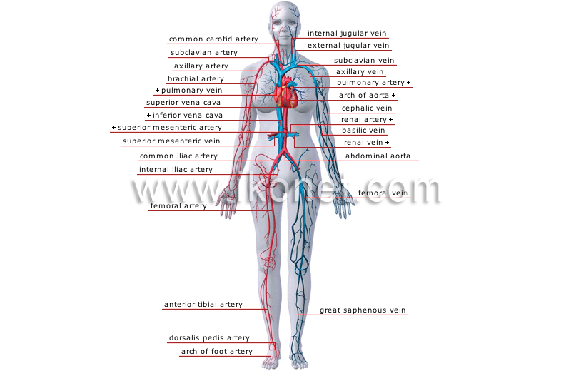
Foot Vein Anatomy
The foots shape along with the bodys natural balance keeping systems make humans capable of not only walking but also running climbing and countless other activities. Venous foot pump voiding.
 Foot Medical Study Student Anatomy Model Showing Bones Toes
Gait at the beginning of a step the distal calf pump is activated.

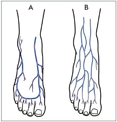
Foot vein anatomy. These foot perforator veins are split into two well separated functional units medial and lateral connected to each plantar vein. Anatomy of the foot perforator veins. I have severe pain in my left foot on the left side outside of foot.
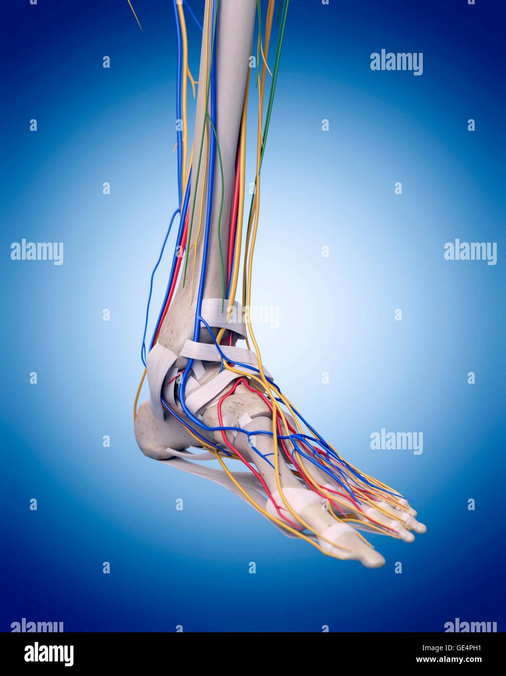
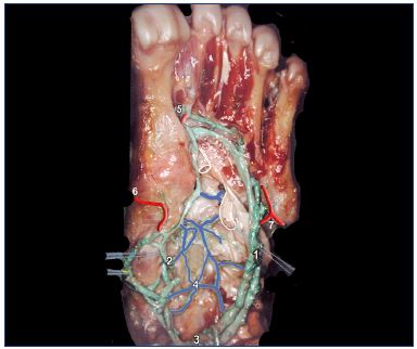
It is accompanied by the dorsalis pedis vein. Foot perforator veins provide direct connections between the plantar veins and the roots for both saphenous systems. There are medial and lateral marginal veins which drain both of the dorsal and plantar parts of the specific sides within the dorsal venous arch alongside the foot.
My foot veins usually pop out but recently they have been flat. Medically reviewed by healthline medical team on april 13 2015. This process is initiated by dorsiflexion of the foot as the leg is lifted to take a step.
Varicose veins are a common pathology seen in the veins of the foot and ankle. One common problem is varicose veins. Superficial vein tributaries drain blood into the dorsal venous arch on the dorsum of the foot at the level of the proximal head of the metatarsal bones.
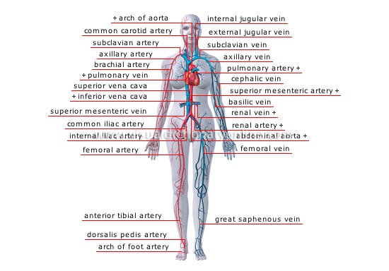
Top 20 doctor insights on. The medial and lateral end of this arch continues through the medial and. The veins of the foot circulate oxygen depleted blood from the tissues back to the heart.
I have sharp stabbing intermitent night pain on top of my left foot left side middle. Figure 27 superficial and perforating veins of the foot and ankle. It interacts along with proximally situated dorsal venous network and receives the dorsal digital as well as dorsal metatarsal veins.
They are ectatic tortuous vessels of the superficial venous system that are at least 3 mm in size that arises from the failure of venous valves to close properly to allow the backward flow of blood. The foot is the lowermost point of the human leg. The anterior compartment muscles contract dorsiflect the foot and empty its veins ie the anterior tibial veins.
Foot veins anatomy 1. Circulation problems of the foot are common in both the elderly and obese people as well as those who stand for long periods of time.
 Veins Arteries Foot Stock Photos Veins Arteries Foot Stock
 Vein Wikipedia
![]() Great Saphenous Vein Anatomy And Clinical Conditions Kenhub
Great Saphenous Vein Anatomy And Clinical Conditions Kenhub
 Figure 2 From The Anatomy And Physiology Of The Venous Foot
 The Venous System Of The Foot Anatomy Physiology And
 Antique
Medically Accurate Anatomy Illustration Of Human Feet Legs
 Thumb Head And Neck Anatomy Vein Human Body Veins
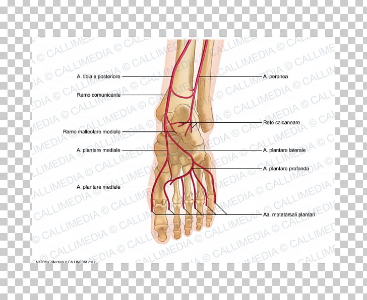
 Anatomy Of Foot And Ankle Perforator Veins Servier
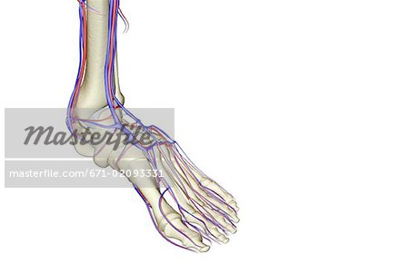 The Blood Supply Of The Foot Stock Photo Masterfile
 7 Anatomy Of The Leg And Dorsum Of The Foot
 Ankle Foot Anatomy
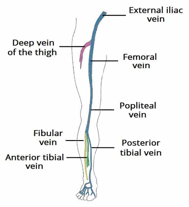
 Leg Vein Map Arterial And Venous Circulation Of The Legs
 Great Saphenous Vein Wikipedia
 Thumb Foot Artery Human Leg Vein Png Clipart Abdomen
![]() Foot Diagram Human Circulatory System Art Print
Foot Diagram Human Circulatory System Art Print
![]() Veins Of The Lower Limb Anatomy Kenhub
Veins Of The Lower Limb Anatomy Kenhub
 Foot Human Leg Common Digital Veins Great Saphenous Vein Png


 Venous Drainage Of The Lower Limb Teachmeanatomy
 The Hemodynamics And Diagnosis Of Venous Disease Sciencedirect
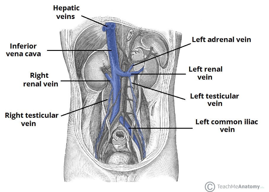 Venous Drainage Of The Abdomen Teachmeanatomy
 Koori Pressure Point Arts
![]() Veins Of The Lower Limb Anatomy Kenhub
Veins Of The Lower Limb Anatomy Kenhub
Belum ada Komentar untuk "Foot Vein Anatomy"
Posting Komentar