Palm Of Hand Anatomy
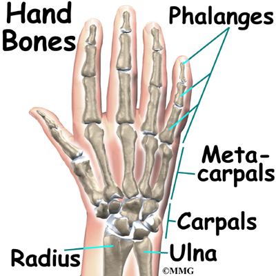
The palm includes five metacarpals and each finger except the thumb contains one proximal phalanx one middle phalanx and one distal phalanx. Also known as the broad palm or metacarpus it consists of the area between the five phalanges finger bones and the carpus wrist joint.
Extensor tendons of the fingers which attach to the middle and distal phalanges and extend.
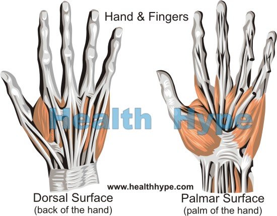
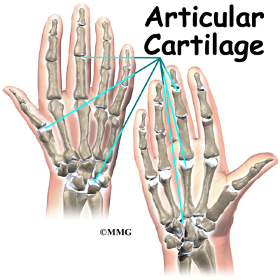
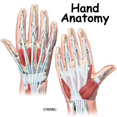
Palm of hand anatomy. The bones in the fingers and thumb are known as phalanges. All small carpal bones join the bones lying next to them and thus make the anatomy of the human hand very complex. The red lines show where the tendons attach the muscles to the bones.
The opisthenar area dorsal is the corresponding area on the posterior part of the hand. Areas of the human hand include. The back of the hand is called the dorsal side.
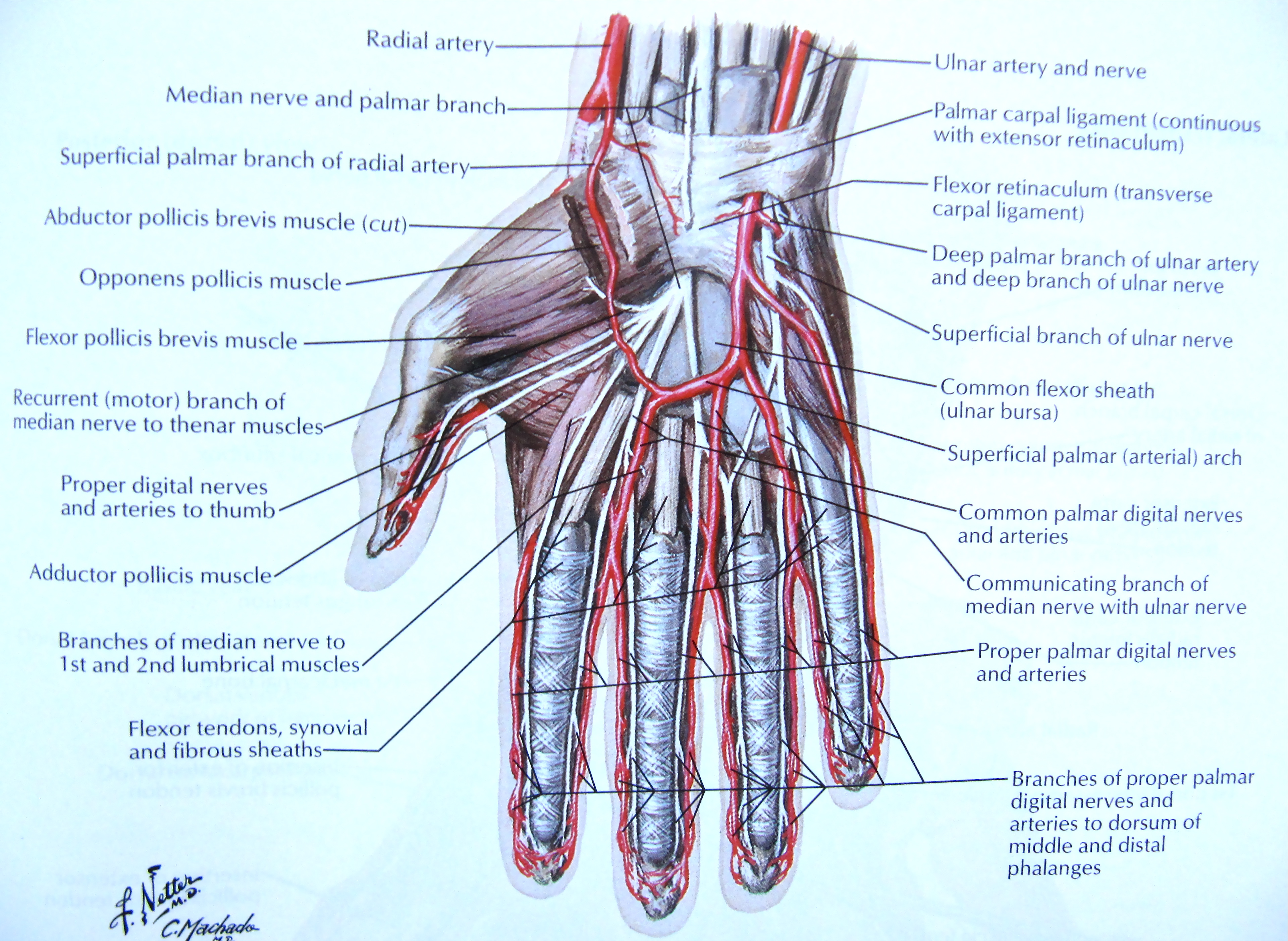
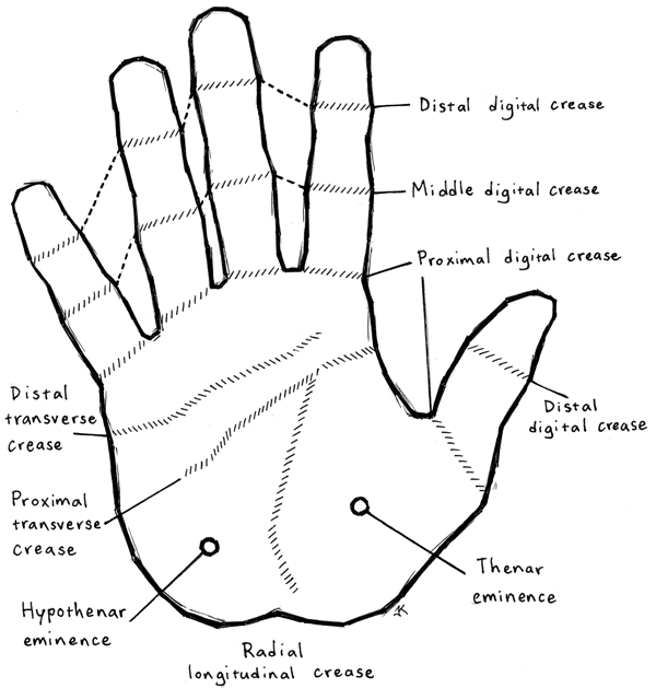
Profundus tendons which pass through the palm side of the wrist and hand. The heel of the hand is the area anteriorly to the bases of the metacarpal bones. The palm volar which is the central region of the anterior part of the hand.
A thickening of the deep fascia covering the palm of the hand. The important structures of the hand can be divided into several categories. Originates from the palmar aponeurosis and flexor retinaculum attaches to the dermis of the skin on the medial margin of the hand.
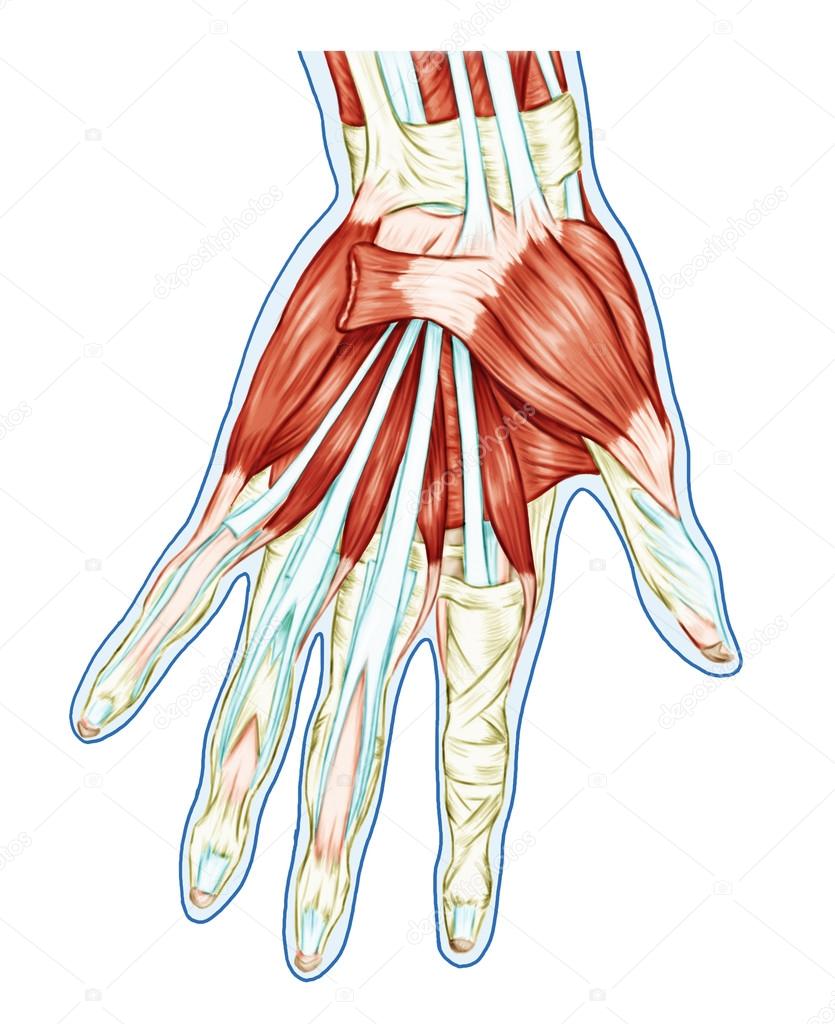
The muscles in the forearm and palm thenar muscles all work together to keep the wrist and hand moving stable and well aligned. Many of the muscles that move the fingers and thumb originate in the forearm. Palmar aponeurosis is composed of very dense connective tissue that extends out into each of the fingers.
The front or palm side of the hand is referred to as the palmar side. Other muscles in the palm attachments. Each hand consists of 19 bones.
Located in the palm are 17 of the 34 muscles that articulate the fingers and thumb and are connected to the hand skeleton through a series of tendons. Wrist palm and fingers are thus made up of several small joints. Wrinkles the skin of the hypothenar eminence and deepens the curvature of the hand improving grip.
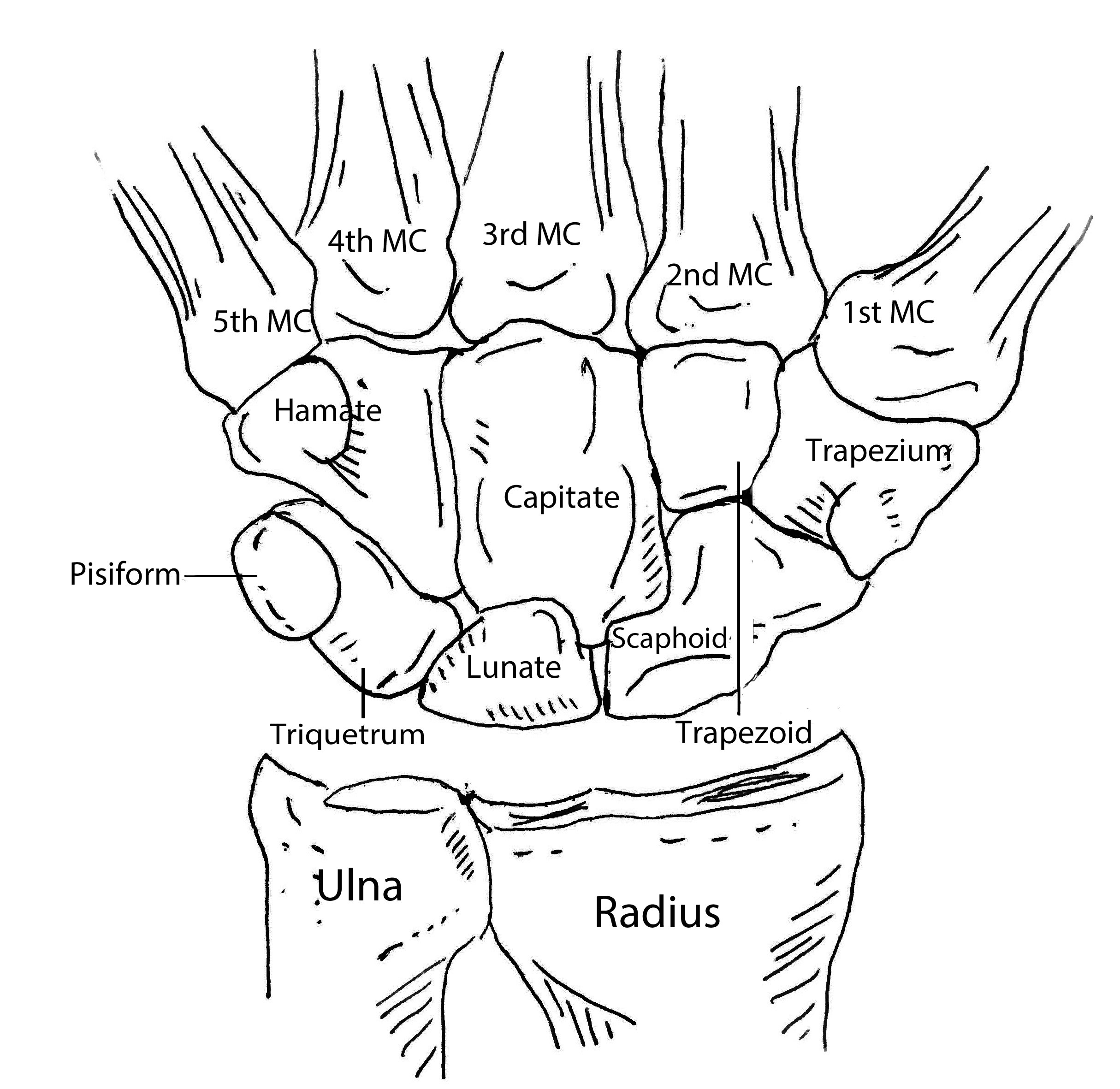
All five metacarpals together are recognized as the metacarpus. The image below shows the bones of the hand from the back side. The main tendons of the hand are.
Superficialis tendons which pass through the palm side of the wrist and hand. The palm comprises the underside of the human hand.
 Hand Bones And Wrist Bones Mnemonics Anatomy And Physiology
 Anatomy Of Muscular System Hand Palm Muscle Tendons
 Superficial Dissection Of The Palm Anatomy Median Nerve
 Dissection Of The Hand Superficial Muscles And Tnedons In The
 Hand Pain Causes Of Pain In The Palm And Back Of Hand
 Hand Anatomy Overview Bones Blood Supply Muscles Geeky


 Finger Hand Palm Image Png 480x640px Finger Anatomy Arm
 Hand And Wrist Injuries Part I Nonemergent Evaluation
 Common Hand And Wrist Conditions Pro Sports Orthopedics
 Muscles Of The Palm Hand For Anatomy Education Stock Image
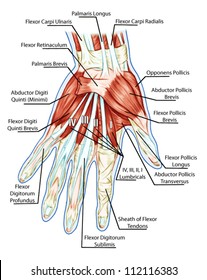 Hand Tendon Images Stock Photos Vectors Shutterstock
 Dupuytren S Recurrence Flexor Muscles And Tendons Of Left
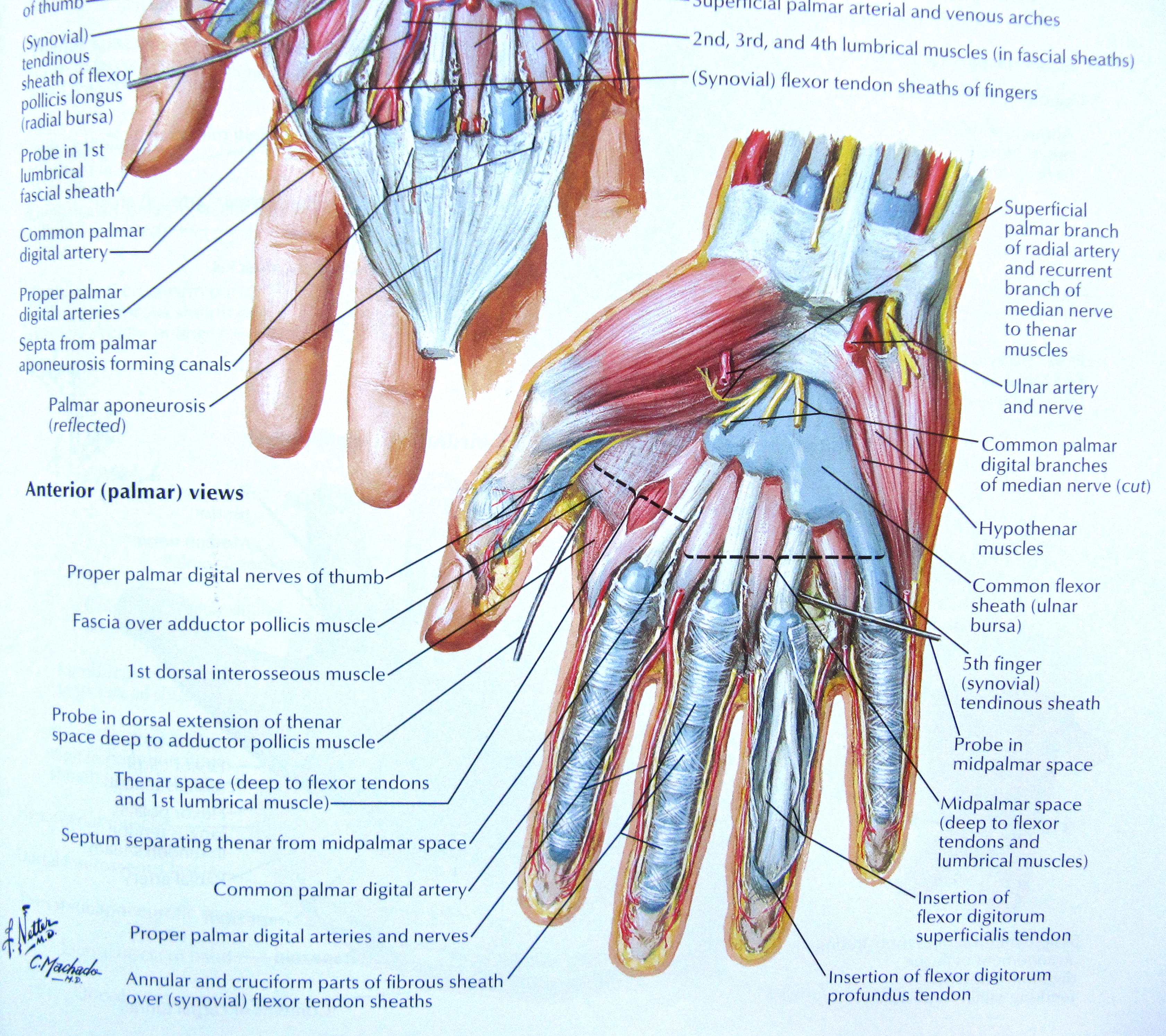 Notes On Anatomy And Physiology The Hand And The Tiger S
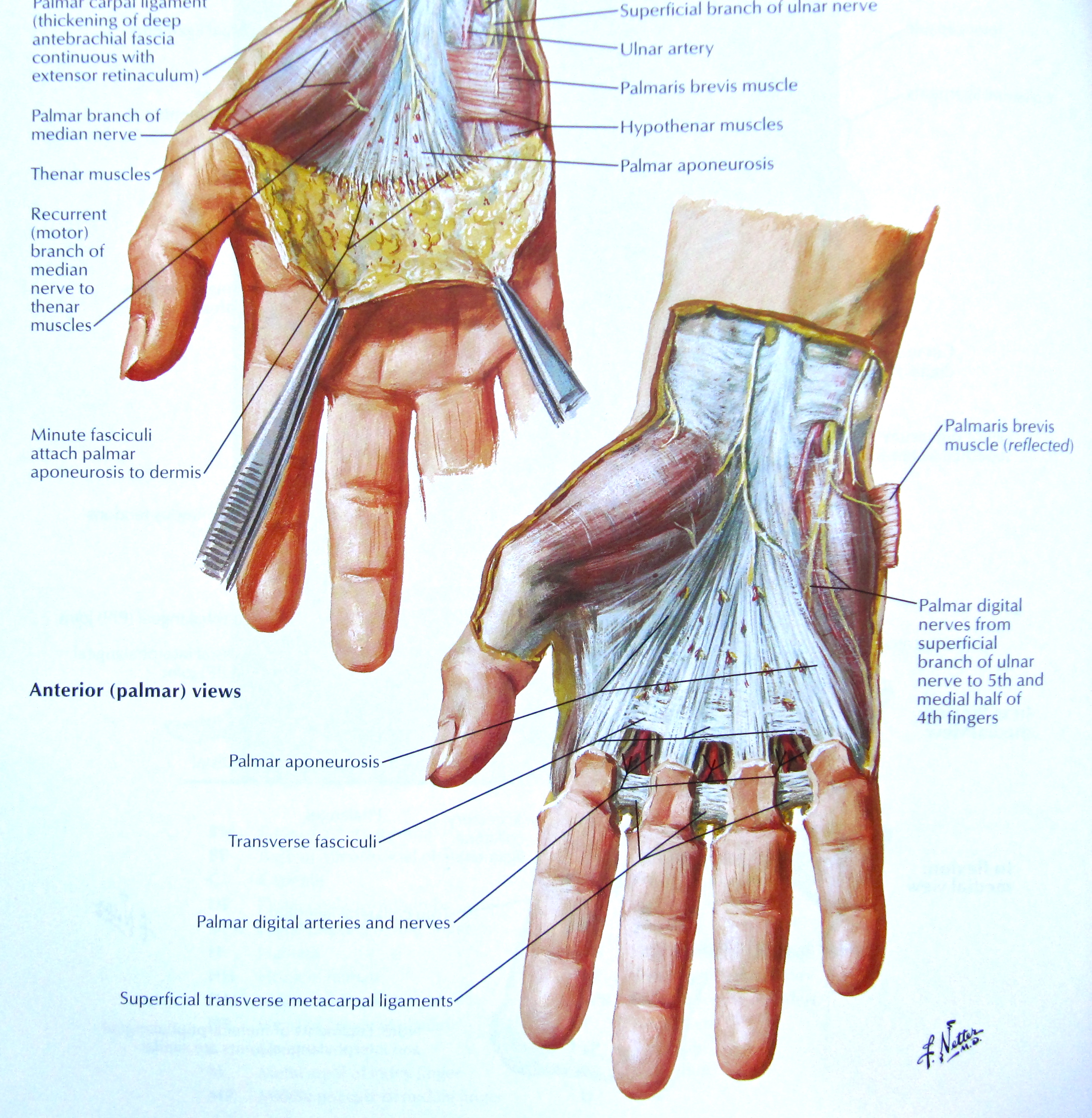 Notes On Anatomy And Physiology The Hand And The Tiger S
 Anatomy Of Muscular System Hand Forearm Palm Muscle
Applied Anatomy Of The Wrist Thumb And Hand
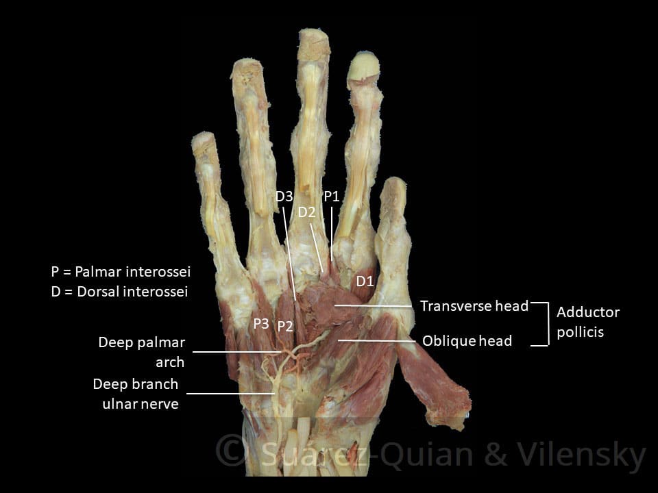 The Muscles Of The Hand Thenar Hypothenar Teachmeanatomy
Anatomy 9 Palm Of Hand Msk Ii Flashcards Memorang
 Notes On Anatomy And Physiology The Hand And The Tiger S
 Anatomy Of Hand Wrist Bones Muscles Tendons Nerves
 Muscles Of The Palm Hand For Anatomy Education Human Physiology
 The Grip Problem Jordan Feigenbaum

 A Guide To Palm Reading Lines Diagram Real Simple


Belum ada Komentar untuk "Palm Of Hand Anatomy"
Posting Komentar