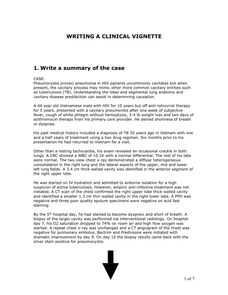
Segmental Lung Anatomy
Each segment is supplied by its own bronchus which is called a segmental bronchus. As long as the surgeons efforts were confined to resection of a lobe or of a whole lung the importance of segmental anatomy was not fully appreciated.
 Anatomy Radiology Key
Removal of a bronchopulmonary segment is known as segmentectomy.

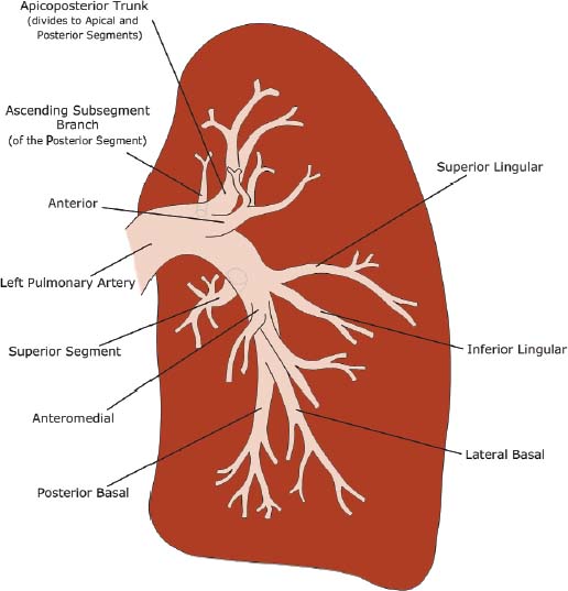
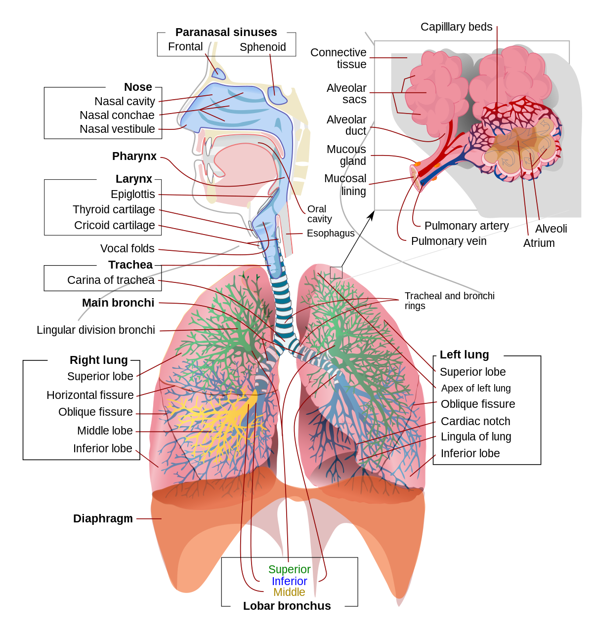
Segmental lung anatomy. These arteries branch from the pulmonary and bronchial arteries and run together through the center of the segment. Segmental and subsegmental pulmonary arteries vary considerably in the location of their origins in whether they arise as common trunks with other arteries or as separate arteries and in their number. It is only meant to simplify the anatomic relation of segments to one another within each lobe and not their exact anatomic locations.
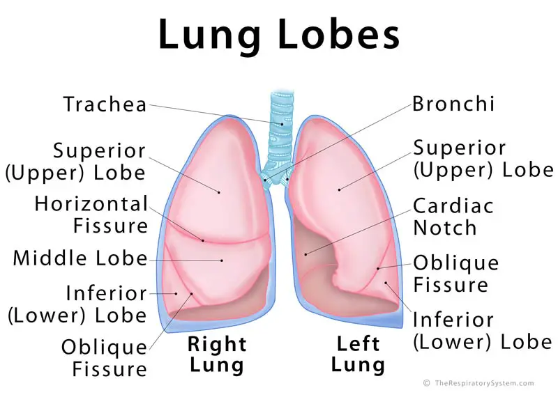
The lingula on the left is part of the left upper lobe and is the equivalent. With this basic symmetric anatomy shared between the lungs there are a few differences that can be described. Knowledge of segmental anatomy is essential for the bronchoscopist who wants to accurately describe the location of endobronchial lesions.
You should be familiar with this anatomy already. In general each lung has 10 segments. Veins and lymphatic vessels drain along the edges of the segment.
Medial and lateral easy to remember m iddle l obe. Each lung is divided into lobes and each lobe is divided into segments. The bronchopulmonary segment is a functionally and anatomically discrete portion of lung supplied by its own segmental bronchus and artery.
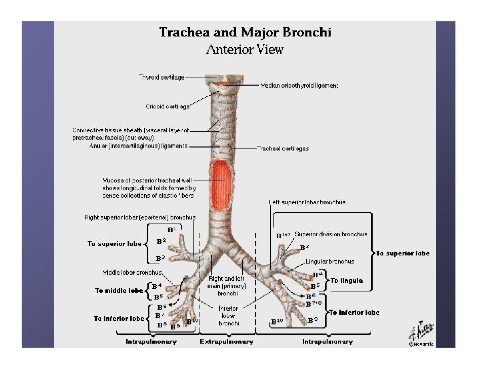
The right lung has 3 lobes divided into 10 segments. The upper lobes contains 3 segments the middle lobe lingula 2 and the lower lobes 5. But when churchill in 1939 successfully removed a segment of the lower lobe for bronchiectasis a new emphasis was created.
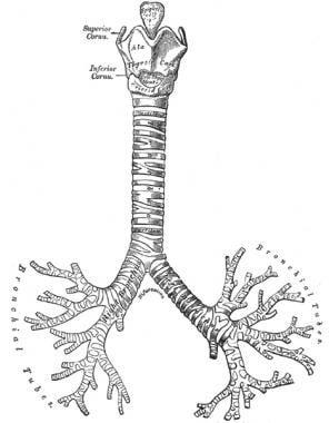
We will learn the anatomy of the tracheobronchial tree and the segments together. There is some form of segmental symmetry between the right and left lungs even though the left lung is smaller and only contains two lobes. The middle lobe on the right has 2 segments.
A bronchopulmonary segment is a portion of lung supplied by a specific segmental bronchus and arteries. The right upper lobe has apical posterior and anterior segments middle lobe has medial and lateral segments and the lower lobe has superior apical and 4 basal segments anterior medial posterior and lateral. Knowledge of bronchopulmonary segment anatomy is important for understanding and interpreting radiographs and other medical images of the lungs especially when needed as reference for surgical resection of diseased lung segments.
1 study figure s1 1. This diagram may help simplify the segmental anatomy of the lung and the pulmonary arteries.
 Ct Pulmonary Angiography
 Lung Lobes Definition Anatomy Functions Picture
 Pleural Cavity Atlas Of Anatomy
 Lung Anatomy Overview Gross Anatomy Microscopic Anatomy

 Lung Scan Radionuclide Venography For Msc 2017
 Bronchopulmonary Segmental Anatomy Radiology Reference
 Pin By Majeja84 On Radiologija Lung Anatomy Anatomy
Tracheobronchial Branching Abnormalities Lobe Based
 Bronchial Anatomy
 Segmental Bronchus Anatomy Segments Of The Lung Images
 Lung Anatomy
 Anatomy Of The Thorax Ct
Ct Pulmonary Angiography
 Writing A Clinical Vignette Write A Summary Of The Case
 Lobar And Segmental Lung Anatomy On Ct
 Bronchus Wikipedia
 5 Chest Radiology Review Manual Dahnert Radiology
 Pulmonary Artery Wikipedia
 Lymph Node Lung Cancer Staging Lung Cancer Staging Anatomy
 Segmental Anatomy Of Lungs Anatomy Of Mediastinum And
 Bronchopulmonary Segmental Anatomy Radiology Reference
 Scheme Of The Bronchial Nomenclatures Each Segmental Node


 The Lungs Anatomy And Physiology Ii
Belum ada Komentar untuk "Segmental Lung Anatomy"
Posting Komentar