Septa Anatomy
Orbital septum a palpabral ligament in the upper and lower eyelids. Biology the plural of septum.
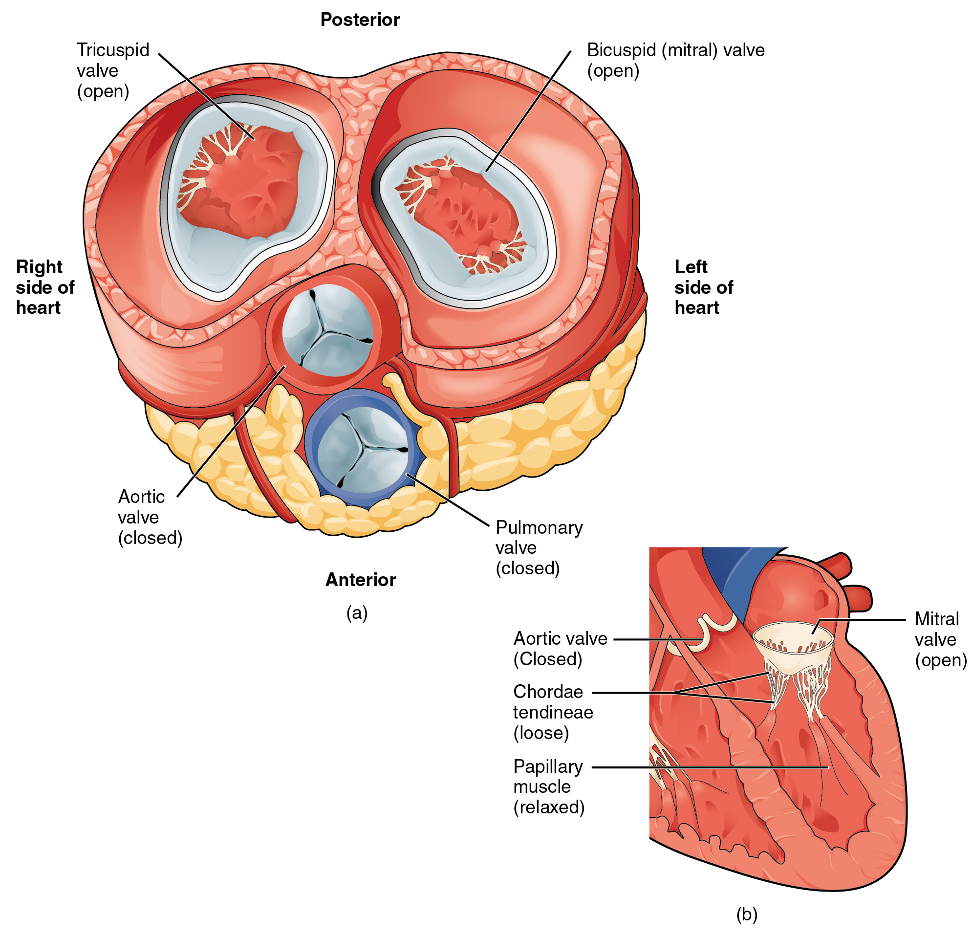
Internal Structure Of The Heart Contemporary Health Issues
Human anatomy alveolar septum.

Septa anatomy. The thin wall which separates the alveoli from each other in the lungs. The septa are simple in some species as in the nautilus fig. The lower chambers the ventricles are separated by the interventricular septum.
Mechanical engineering the plural of septum. Southeastern pennsylvania transportation authority serving bucks chester delaware montgomery and philadelphia counties. Uterine septum a.
The septa are physical extensions of the myocardium lined with endocardium. Septa is a plural form of septum general anatomy. The septa may act as a barrier or conduit for the spread of pus blood urine and neoplasms in the perinephric space.
Underwood an anatomist at kings college in london. The presence of septa at or near the floor of the sinus are. The ventricles in turn pump blood to the lungs and to the remainder of the body.
The atria receive blood from various parts of the body and pass it into the ventricles. Southeastern pennsylvania transportation authority serving bucks chester delaware montgomery and philadelphia counties. Anatomy the plural of septum.
A dividing partition between tissues or cavities. Often also some other septa are developed so that each tangential tube seems to be composed of four to six joints or segments. Septa mordane susan brown is a teacher to sansa and arya stark maisie williams.
The word septum is derived from the latin for something that encloses in this case a septum plural septa refers to a wall or partition that divides the heart into chambers. Their septa are fine and barely or not at all denticulated. Perinephric bridging septa or septa of kunin are composed of numerous fibrous lamellae which traverse the perinephric fat 12 where they suspend the kidneys within the perirenal space.
Septum pellucidum or septum lucidum a thin structure separating two fluid pockets in the brain. In anatomy underwoods septa or maxillary sinus septa singular septum are fin shaped projections of bone that may exist in the maxillary sinus first described in 1910 by arthur s. A partition known as the interatrial septum.
 Alveolar Septum An Overview Sciencedirect Topics
 Anatomy And Physiology Of The Male Reproductive System
 High Resolution Ct Of The Lung
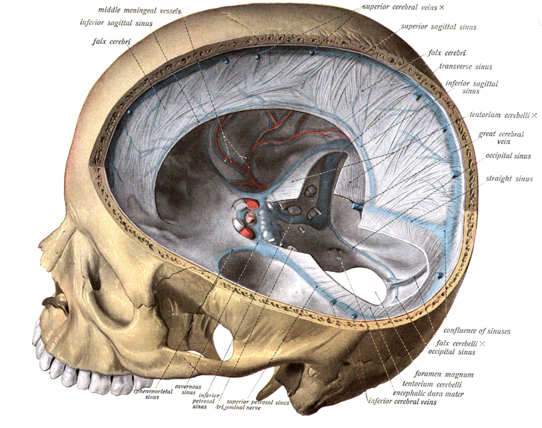
 Cranial Dura Septa And Dural Venous Sinuses Mid Sagittal
 The Location And Orientation Of Maxillary Sinus Septa Mss
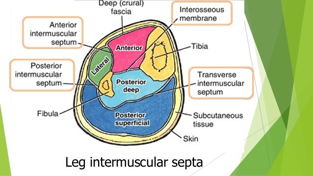
![]() Intermuscular Septa Stock Photos Intermuscular Septa Stock
Intermuscular Septa Stock Photos Intermuscular Septa Stock
 19 1 Heart Anatomy Anatomy And Physiology
 Table 8 Cranial Meninges And Spaces Cranial Dural Septa
 The Muscles And Fasciae Of The Arm Human Anatomy
 Dural Septa And Dural Venous Sinuses A Dural Septa Are
 Ultrastructural Anatomy Of A Lacrimal Lobule 1 Interlobular
 Meninges Overview On The Structure Of The Individual Layers
 Cranial Dural Septa 12 Motion Sickness Viii X Anatomy
 Dural Septa And Sinuses Youtube
 An Atlas Of Human Anatomy For Students And Physicians
:background_color(FFFFFF):format(jpeg)/images/library/11260/anatomy-testis-and-epididymis_english__1_.jpg) Testes Anatomy Definition And Diagram Kenhub
Testes Anatomy Definition And Diagram Kenhub
 Interlobular Septa Radiology Reference Article
What Does The Fascia System Do Quora
 Serous Salivary Gland Anatomy Physiology Wikivet English
 Duplication And Septa Of The Bladder


Belum ada Komentar untuk "Septa Anatomy"
Posting Komentar