Anatomy Of The Index Finger
A digit includes the hand bones but these bones are not separated into individual appendages like a finger. Picture of finger anatomy.
 Surgical Fixation Of The Left Index Finger Medical
Ligaments connect finger bones and help keep them in place.

Anatomy of the index finger. Fingers have a complex anatomy. The phalanges singular phalanx the 14 narrow bones that make up the fingers of each hand. The median nerve innervates the fingers skin.
The index middle ring and fifth digits have proximal middle and distal phalanges and three hinged joints. Each finger has three phalanges the distal middle and proximal. The thumb has two.
The little finger and index finger both have an extra muscle. There are no muscles in the fingers. Distal interphalangeal dip proximal interphalangeal pip and metacarpophalangeal mcp.
And fingers move by the pull of forearm muscles on the tendons. The thumb has two. The index finger has three phalanges.
It is also called the index finger or the forefinger. The thumb has two of each. The index finger is composed of three bones.
This finger often possesses the largest amount of sensitivity and greatest dexterity of any of the fingers. The index finger does not contain any muscles but is controlled by muscles in the hand by attachments of tendons to the bones. Each finger has 3 phalanges bones and 3 hinged joints.
Oxygenated blood arrives at the finger through the common palmar artery which extends off of the palmar arch connecting the ulnar and radial arteries. Each finger has three phalanges the distal middle and proximal. Fingers are constructed of ligaments strong supportive tissue connecting bone to bone tendons attachment tissue from muscle to bone and three phalanges bones.
The thumb has a distal and proximal phalanx as well as an interphalangeal and mcp joint. The distal phalanx intermediate phalanx and proximal phalanx. Anatomy of the fingers the human finger is mainly a bony structure with multiple joints giving it strength and flexibility.
Tendons connect muscles to bones. Finger movement is controlled by muscles in the forearms that pull on finger tendons. Basic anatomy of the finger.
The extensor indicis extends the index finger while the palmar interosseus adducts it.
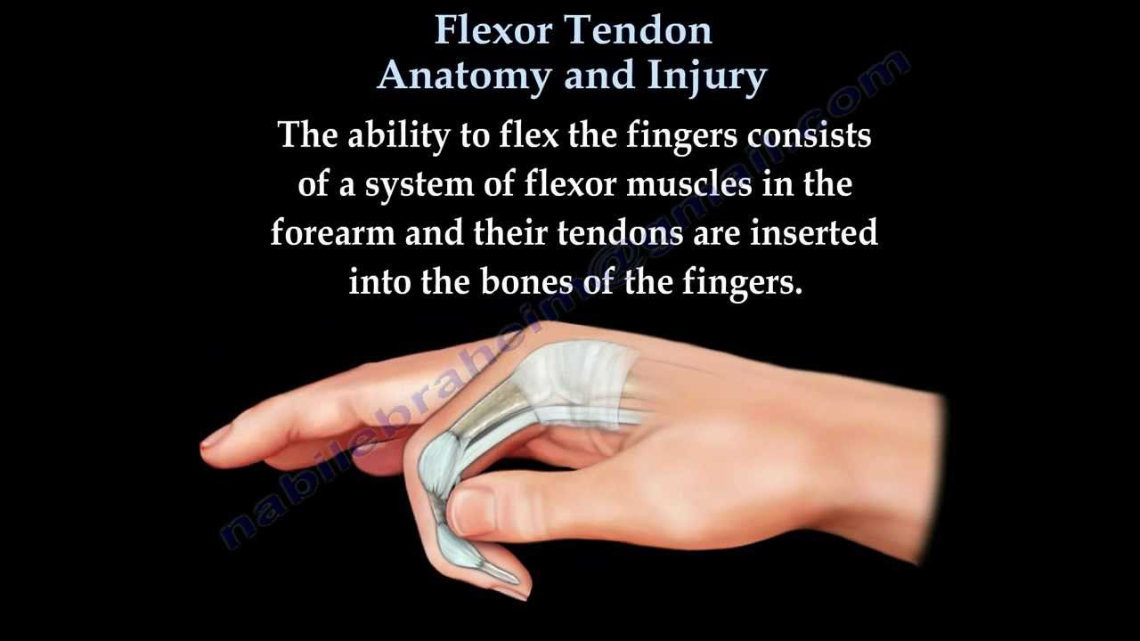 Flexor Tendon Anatomy And Injury Everything You Need To Know Dr Nabil Ebraheim
Trigger Finger Trigger Thumb Orthoinfo Aaos
 Ucsd S Practical Guide To Clinical Medicine
 Jpma Journal Of Pakistan Medical Association
 What Peculiarities Of The Anatomy Of Hands Should Be Taken
 Human Anatomy Figure 9 4

 Fingertip Amputations Finger Flaps Hand Orthobullets


 Right Index Finger Fractures Medical Illustration Human
 Diagram Of The Fingers Reading Industrial Wiring Diagrams

 Related Image Hand Anatomy Anatomy Illustration
 The Muscles And Fasciae Of The Hand Human Anatomy

 Metacarpals Approach Dorsal To 2nd Metacarpal Shaft
Plos One Biomechanical Constraints Underlying Motor
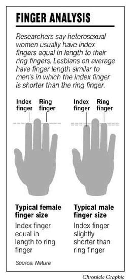 Finger Length Points To Sexual Orientation Anatomy Quirk
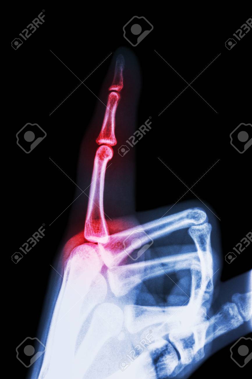 Rheumatoid Arthritis Gouty Arthritis Film X Ray Index Finger
 Acute Finger Injuries Part I Tendons And Ligaments
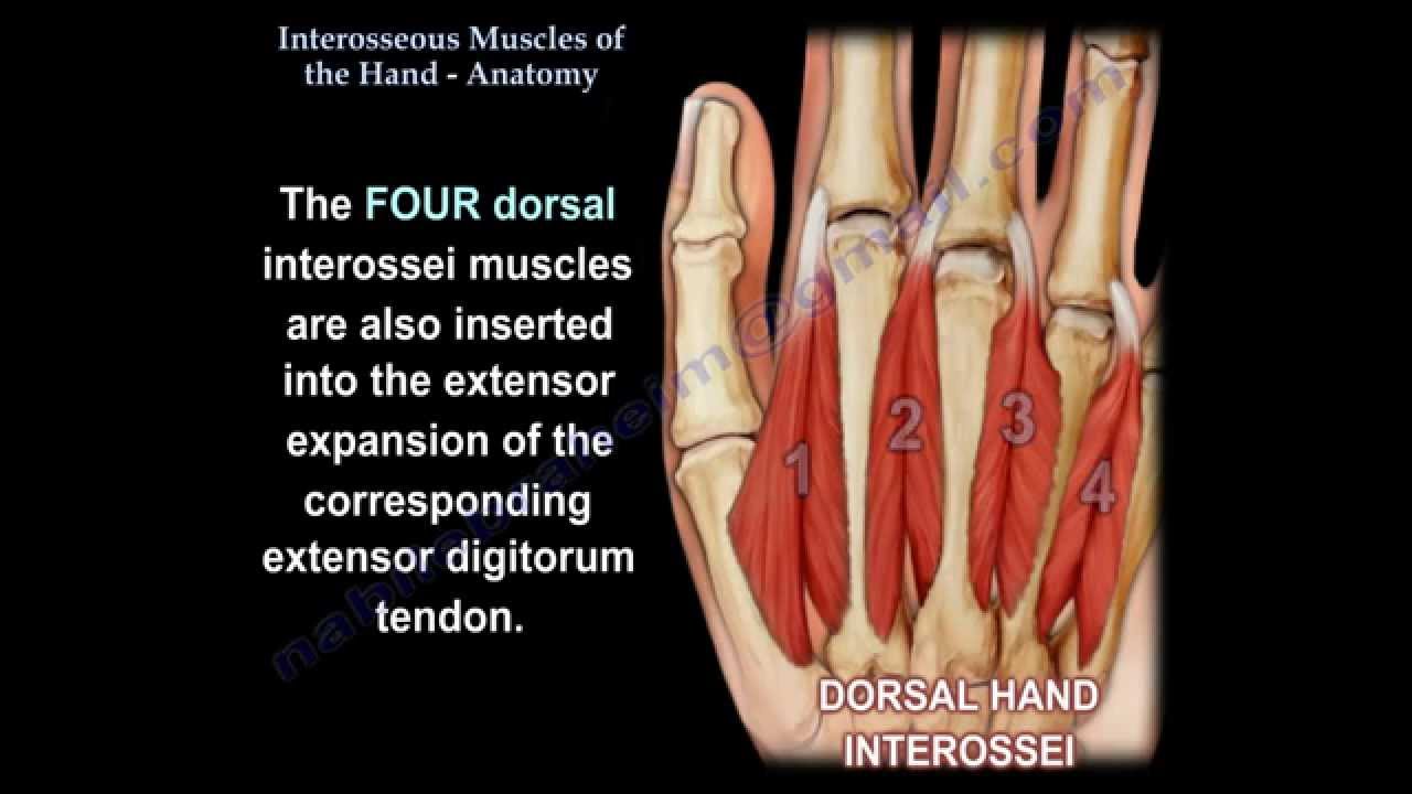 Interosseous Muscles Of The Hand Anatomy Everything You Need To Know Dr Nabil Ebraheim
 Anatomy Of Carpal Tunnel Syndrome Everything You Need To Know Dr Nabil Ebraheim
 Describing Where Things Are On The Hand Sketchy Medicine
Belum ada Komentar untuk "Anatomy Of The Index Finger"
Posting Komentar