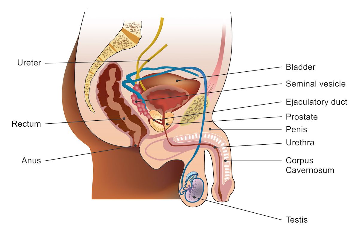
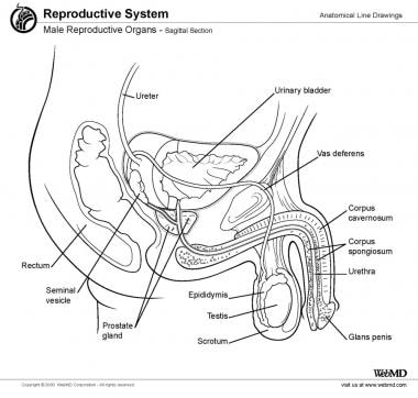
Anatomy Of Testis
It is homologous to the female ovary. Whereas sperm production is controlled both by the anterior pituitary follicle stimulating hormone and gonadal testosterone.
![]() Diagram Pictures Testis And Epididymis Anatomy Kenhub
Diagram Pictures Testis And Epididymis Anatomy Kenhub
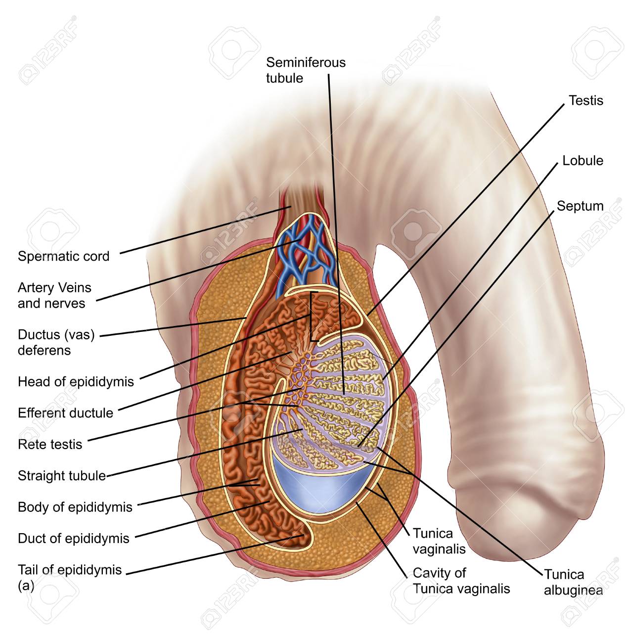
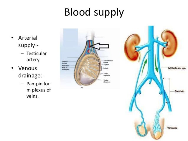
They are suspended from the abdomen by the spermatic cord collection of vessels nerves and ducts that supply the testes.

Anatomy of testis. Testis plural testes also called testicle in animals the organ that produces sperm the male reproductive cell and androgens the male hormones. Benninghoff 1993 b. Spermatic cord and vascular supply of the testis.
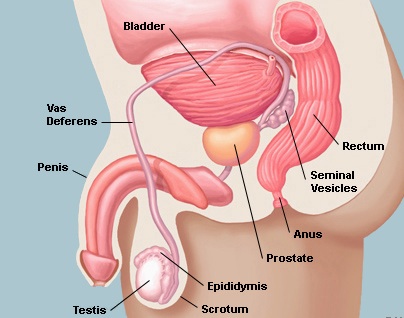
Testicle or testis is the male reproductive gland or gonad in all animals including humans. Anatomy of the scrotum. The epididymis is 510 mm thick and extends from the upper to.
Plz like and subscribe my channelanatomy of testis. Detail anatomy on small intestine its anatomy its part its physiology. Sahar hafeez videos done by.
The testes are responsible for the production of sperm cells and the male sex hormone testosterone. In humans the testes occur as a pair of oval shaped organs. Commonly the left testicle lies lower than the right.
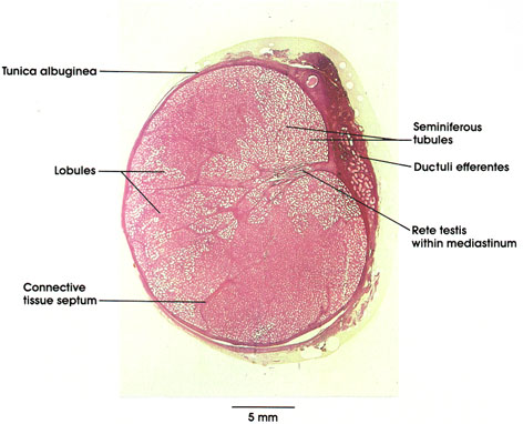
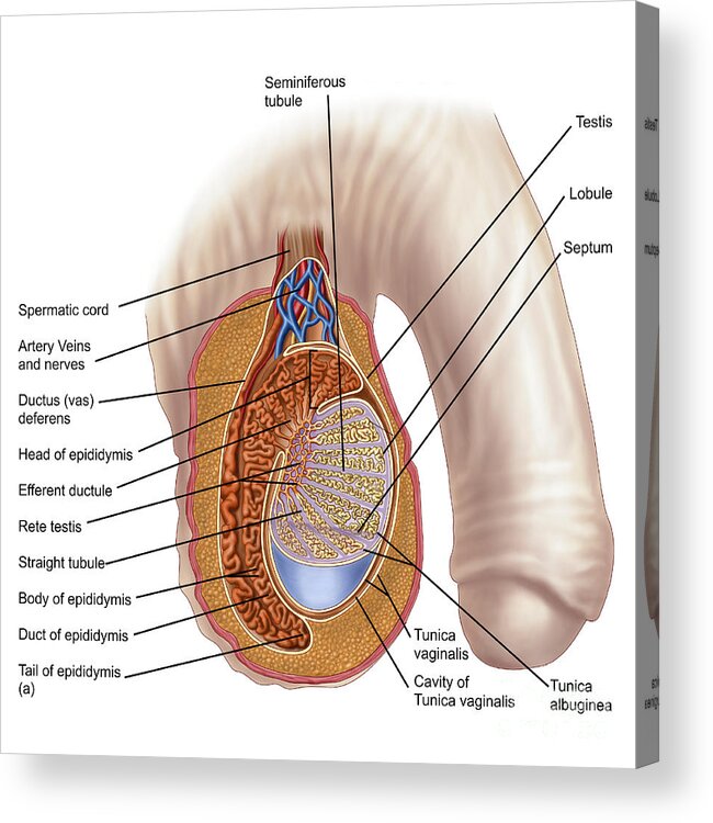
A dense capsule encasing each testis. Scrotum is a cutaneous skin sac that protects the testes. Anatomy of the testis.
Welcome to a series of educational videos covering the regional anatomy of human body. Scrotum and coverings of the testes. Testis commonly known as the testicles are a pair of ovoid glandular organs that are central to the function of the male reproductive system.
On the testis we can observe two sides. The functions of the testes are to produce both sperm and androgens primarily testosterone. Project designer presenter.
Testosterone release is controlled by the anterior pituitary luteinizing hormone. The testes are located within the scrotum with the epididymis situated on the posterolateral aspect of each testicle. The tubules are surrounded by the connective.
After sperm is created in the seminiferous tubules. Inhibits direct extension of tumor. Gross anatomy of the testis anatomy of the epididymis.
The following structures enclose the contents of the scrotum. The intrauterine development of the testes occurs retroperitoneally. They are contained within the scrotal sac which is located directly behind the penis and in front of the anus.
The efferent ducts are a series of tubes that join the rete testis to. Seminiferous tubules are coiled tubes that make up most of each testis. Anatomy and function of testes seminiferous tubules.
Each testicle is considered a separate site unless bilateral involvement is stated to be metastatic from one side to the other.
Anatomy And Physiology Of The Male Reproductive System
 Testicles Facts Function Diseases Live Science
The Male Reproductive System Junqueira S Basic Histology
 Anatomy Of The Testicle
 Anatomy Of The Scrotum And Testes Of Lambs Download
 Anatomy Of Male Testis

 Testes And Epididymis Anatomy Overview Gross Anatomy
 Anatomy Atlases Atlas Of Microscopic Anatomy Section 1 Cells
 Testis Development Embryology And Anatomy Springerlink
 Testis Spermatic Cord
 Scrotal Normal Ultrasoundpaedia
 Testes Anatomy And Function Diagram Conditions And
 The Testicles Canadian Cancer Society
 Figure Testicle Vas Ductus Deferens Head
 Testes Anatomy And Physiology
 Ucsf Department Of Urology Testicular Cancer
 1 Functional Anatomy Of The Testis Medicine Mvst 1b With
 Testis Epididymis
 Anatomy Of Male Testis Acrylic Print

 Testicular Ultrasound Pathology Of The Testes

 Male Testis Anatomy Illustration License Download Or
Belum ada Komentar untuk "Anatomy Of Testis"
Posting Komentar