Anterior Teeth Anatomy
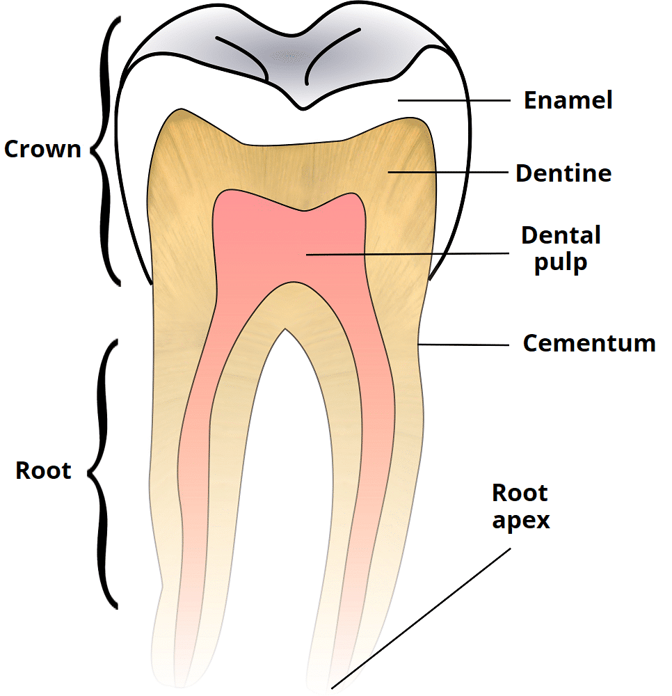
Anterior teeth ordinarily have one root canal. The tip of the root is known as the apex.
The tooth contains an aggregate of blood vessels nerves and cellular connective tissue called the dental pulp.

Anterior teeth anatomy. The dental pulp is housed within a pulp chamber and root canal of a tooth. And incisal edges or single cusped crowns ending in narrow edges. Learn vocabulary terms and more with flashcards games and other study tools.
The development appearance and classification of teeth fall within its purview. The anterior teeth are the teeth that are most visible when a person smiles and they are also among the first baby teeth to be lost and replaced by permanent adult teeth. Internal anatomy of anterior tooth.
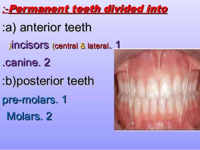
These anterior teeth are wider labiolingually than mesiodistastally mandibular canines these canines have contact areas that are more incisally located when compared with canines from the opposing arch. Anterior teeth include the central and lateral incisors and the canine teeth. Usually there are 20 primary te.
Anterior teeth are located in the anterior part of the jaw. Start studying anatomy anterior teeth. Dental anatomy is also a taxonomical science.
Dental anatomy is a field of anatomy dedicated to the study of human tooth structures. It is concerned with the naming of teeth and the structures of which they are made this information serving a practical purpose in dental treatment. To that part of the arch in which they are located.
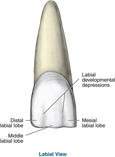
All anterior teeth are composed of four developmental lobes. In cross section canal is ovoid mesiodistally in cervical third rounded in middle third and round in the apical third. Permanent anterior teeth include the incisors and canines figure 16 1 see figure 2 4 15 1.
Tooth formation begins before birth and the teeths eventual morphology is dictated during this time. Just behind them sit the premolars and the molars are located in the very back of the mouth. They make up the six upper and six lower front teeth.
Three labial lobes named mesiolabial middle labial and distolabial and one lingual lobe figure 16 2. Edges are designed to incise bite off relatively large amounts of food in eating. Root canal is broad labio palatally conical in shape and centrally located.
Multiple canals occur in posterior teeth. The eight incisors are the flat squarish sharp teeth located front and center and we use them for cutting food. Root and root canal it has one root with one root canal.
The anterior teeth are the six teeth at the front of the mouth on each jaw.
Finishing Procedures In Orthodontics Dental Dimensions And
 Direct Anterior Restoration Internal Anatomy
 A New System For Classifying Root And Root Canal Morphology
 Anterior Anatomy And The Science Of A Natural Smile
 Microdontia An Overview Sciencedirect Topics
 Comparison Of Selected Anatomical Features Of Permanent
Finishing Procedures In Orthodontics Dental Dimensions And
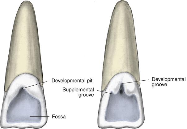 16 Permanent Anterior Teeth Pocket Dentistry
Tooth Anatomy Marginal Ridges Withers Dental
 Teeth Names And Numbers Diagram Names Number And
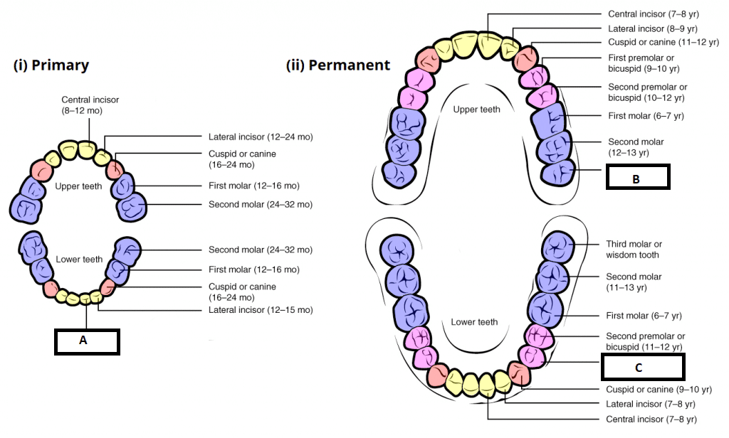 Child And Adult Dentition Teeth Structure Primary
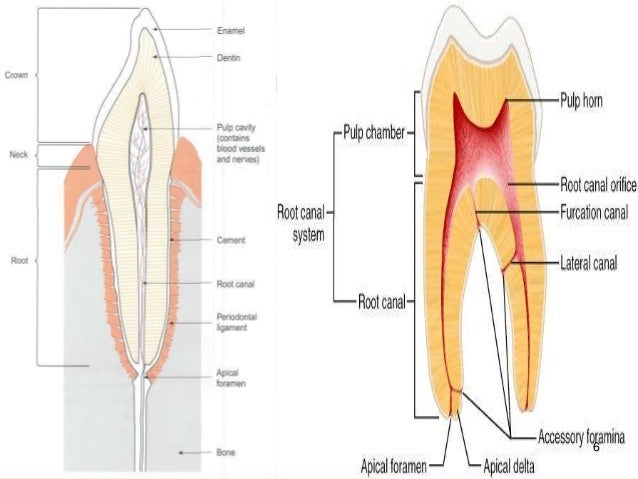 Internal Anatomy Of Anterior Tooth
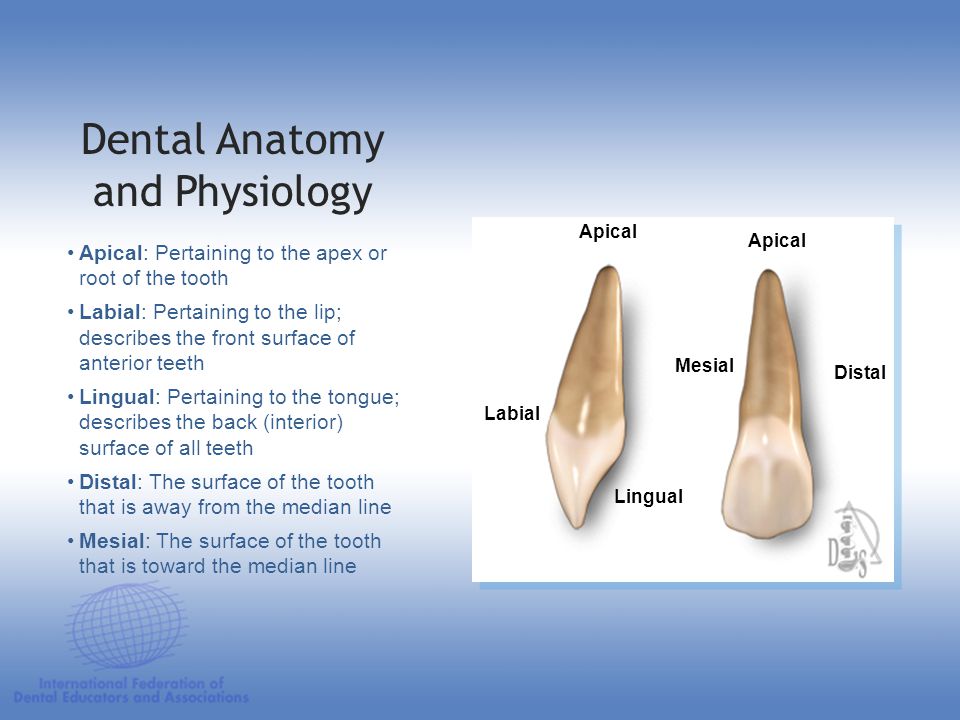 Dental Anatomy Physiology Ppt Download
 Oral Maxillofacial Regional Anesthesia Nysora
 Dr Glaser S 10 Commandments Of Attachment Design
 16 Permanent Anterior Teeth Pocket Dentistry
 Simplifying Anterior Anatomy Glossary
 Learning Anatomy Of The Anterior Teeth Ptc
:watermark(/images/watermark_only.png,0,0,0):watermark(/images/logo_url.png,-10,-10,0):format(jpeg)/images/anatomy_term/cementum/2ViGaJ0wmthQUoI8YSMG3w_Cementum_01.png) Teeth Anatomy Blood Supply And Innervation Kenhub
Teeth Anatomy Blood Supply And Innervation Kenhub
:watermark(/images/watermark_5000_10percent.png,0,0,0):watermark(/images/logo_url.png,-10,-10,0):format(jpeg)/images/atlas_overview_image/678/dScLwAxcZVM6ZiQ0oSXy9A_the-anatomy-of-the-tooth_english.jpg) Teeth Anatomy Blood Supply And Innervation Kenhub
Teeth Anatomy Blood Supply And Innervation Kenhub
 Incisors And Anterior Teeth 1 800 Dentist
 Child And Adult Dentition Teeth Structure Primary
 Dental Anatomy Reference Guide
Introduction To Dental Anatomy Dental Anatomy Physiology
 Glossary Of Technical Terms Clearcorrect Support
 Surfaces Of The Teeth An Overview Of Dental Anatomy
The Primary Deciduous Teeth Dental Anatomy Physiology
 Identifying Anterior Teeth Page 210 In Our Dental Anatomy
 Oral Maxillofacial Regional Anesthesia Nysora



Belum ada Komentar untuk "Anterior Teeth Anatomy"
Posting Komentar