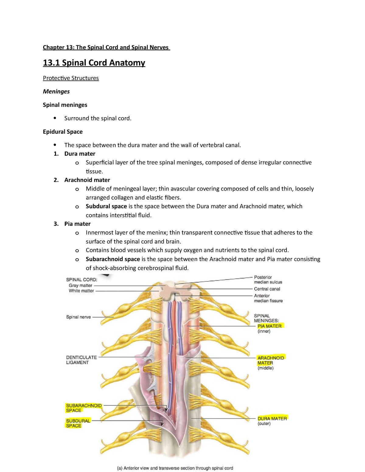
Epidural Space Anatomy
In humans the epidural space contains lymphatics spinal nerve roots loose connective tissue fatty tissue small arteries. These veins are predominantly in the antero lateral part of the epidural space.
 Administering Top Ups Of Epidural Analgesia Nursing Times
The epidural space contains fat epidural veins spinal nerve roots and connective tissue figure 6b the subdural space is a potential space between the dura and the arachnoid and contains a serous fluid.

Epidural space anatomy. Blood vessels these veins communicate with the segmental veins of the neck the intercostal azygos and lumbar veins. It is the space within the canal formed by the surrounding vertebrae lying outside the dura mater which encloses the arachnoid mater subarachnoid space the cerebrospinal fluid and the spinal cord. The boundaries of the epidural space are summarized in table 1 and the definitions of the cervical thoracic lumbar and sacral regions are defined in table 2.
The epidural space contains fat the dural sac spinal nerves blood vessels and connective tissue. The epidural space is the area between the outermost layer of tissue and the inside surface of bone in which the spinal cord is contained ie the inside surface of the spinal canal. Anatomy of epidural space.
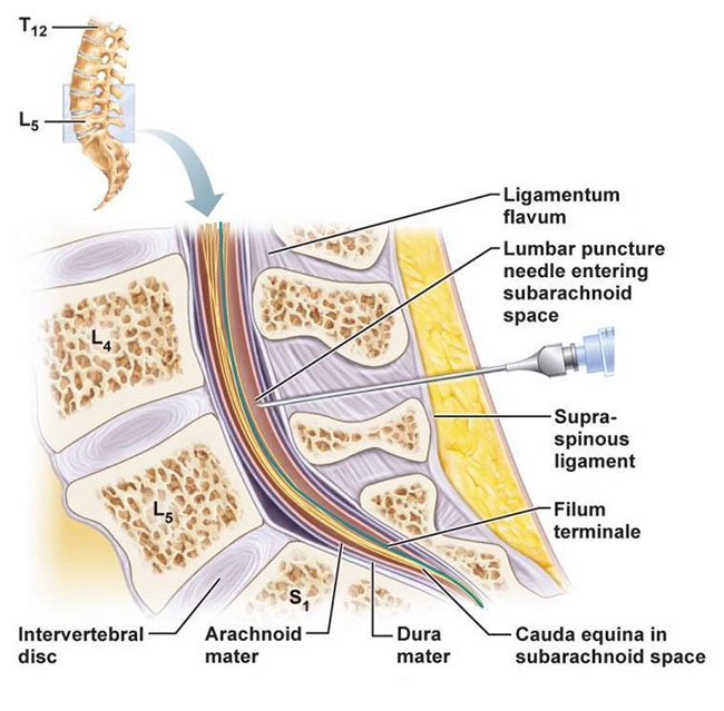
Epidural space subarachnoid space and subdural space anatomy in this image you will find epidural space subdural space subarachnoid space pia mater arachnoid dura mater spinal meninges bone of the vertebra dorsal root ganglion the body of the vertebra in it. The subdural compartment is formed by flat neuroepithelial cells that have long interlacing branches. The spinal epidural space is located in the spinal canal between the spinal dura mater and the vertebral column and extends from the foramen magnum to the sacral canal at the level of s23 3.
The other two spaces are in the spinal cord itself. With the veins of bones of the vertebral column the internal and external vertebral plexuses form batsons plexus. It is typically 4 6 mm in depth 4.
The epidural space runs the length of the spine.
 Neuroanatomy Online Lab 4 External And Internal Anatomy
 Epidural Steroids Home Page The Burton Report
 Chapter 13 Spinal Cord And Spinal Nerves Biol 235 Au
 Anaesthesia Uk Anatomy Relevant To Epidural And
 Vertebral Osteomyelitis Rare Spinal Infection Can Cause
Spinal Injections Orthoinfo Aaos
Epiduroscopic Images Of Spinal Anatomy Neupsy Key

Quantitative Anatomy Of The Thoracolumbar Epidural Space
/epiduralspace-56a05e4f3df78cafdaa149f7.gif) Epidural Space Anatomy And Injections
Epidural Space Anatomy And Injections
 Image 09765 Im03 Cervical Epidural Steroid Injections At C7 T1 Illustration
 Sc Brain Meninges At South College Studyblue

 Applied And Radiological Anatomy Of The Epidural Space For
 Technology Medisight
 Lumbar Spine Anatomy Overview Gross Anatomy Natural Variants
Graphical Representation Of The Epidural Space And
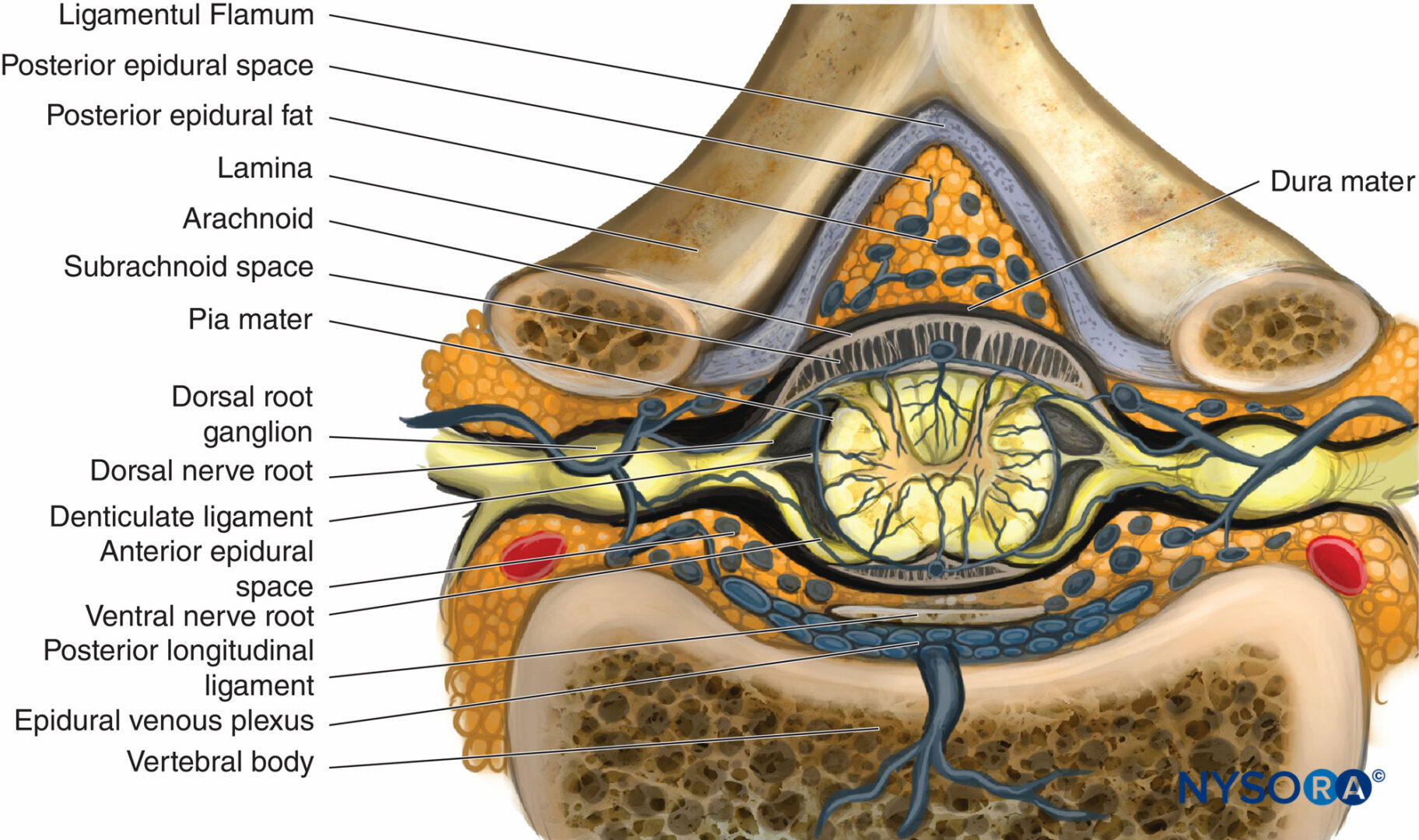
 Neuraxial Anatomy Nysora
 Epidural Anesthesia And Analgesia Nysora
 Epidural Space An Overview Sciencedirect Topics
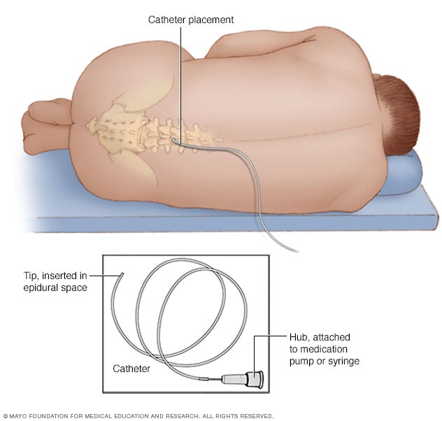 Epidural Delivery Of Pain Medication Mayo Clinic
 Pin On Epidural Space
Belum ada Komentar untuk "Epidural Space Anatomy"
Posting Komentar