Foot Anatomy Mri
Your doctor with the help of a radiologist can then examine these images to determine whether there is anything wrong with your foot or ankle. Unable to process the form.
 Essr 2016 P 0029 Mri At Forefront Of Forefoot Pain Epos
Magnetic resonance imaging otherwise known as mri uses a combination of magnetic fields and radio waves to take images of the internal structures of your body.

Foot anatomy mri. Foot radiograph an approach foot radiographs are commonly performed in emergency departments usually after sport related trauma and often with a clinical request that states lateral border pain. For hind and mid foot a 12 to 14 cm field of view is applied. Anatomy of the ankle and foot mri atlas of the human body using cross sectional imaging.
The exquisite soft tissue contrast resolution noninvasive nature. Often a foot x ray is also requested for the investigation of osteomyelitis arthritides or. Loss of joint alignment can represent severe injury even in the absence of a fracture.
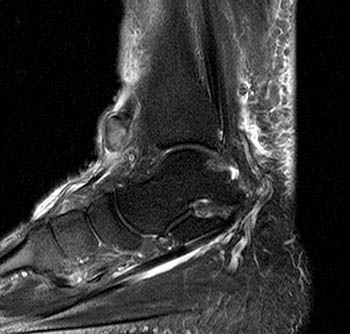
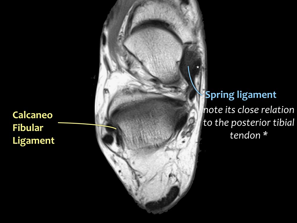
When checking any post traumatic foot x ray it is crucial to assess alignment of the bones at the joints. Depending on the clinical question mri of the foot should be tailored to a hindfoot midfoot or forefoot examination. This webpage presents the anatomical structures found on ankle mri.
Anatomical structures of the ankle and foot and particular regions major joints are visible as dynamic labelled images. Approach to foot series. Foot radiographs are performed for a variety of indications including 1 4.
Mri of the ankle. Click on a link to get sagittal view t1 axial view t2fatsat coronal view t2fatsat sagittal view t2fatsat. For the forefoot a 10 to 12 cm field of view is used to image the smaller peripheral joints in detail.
Knee shoulder shoulder arthrogram ankle elbow. The series is often utilised in emergency departments after trauma or sports related injuries 24. It demonstrates abnormalities in the bones and soft tissues before they become evident at other imaging modalities.
Use the mouse to scroll or the arrows. Magnetic resonance mr imaging has opened new horizons in the diagnosis and treatment of many musculoskeletal diseases of the ankle and foot. Check for errors and try again.
Foot and ankle mri what you should know. The foot series is comprised of a dorsoplantar dp medial oblique and a lateral projection. Remember to check the whole film though.
The cross sectional human anatomic atlas of the ankle and foot is a new tool based on mr images of the human body.
 Musculoskeletal Mri
 Radiologic Evaluation Of The Ankle And Foot Fundamentals
 Figure 1 From Midsagittal Anatomy Of Lumbar Lordosis In

 Mri Ankle Anatomy


 Mri Ankle Anatomy Ankle Anatomy Anatomy Human Anatomy
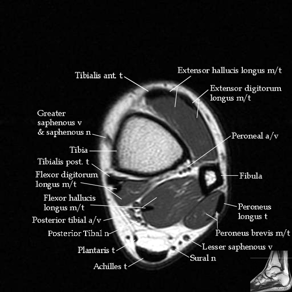
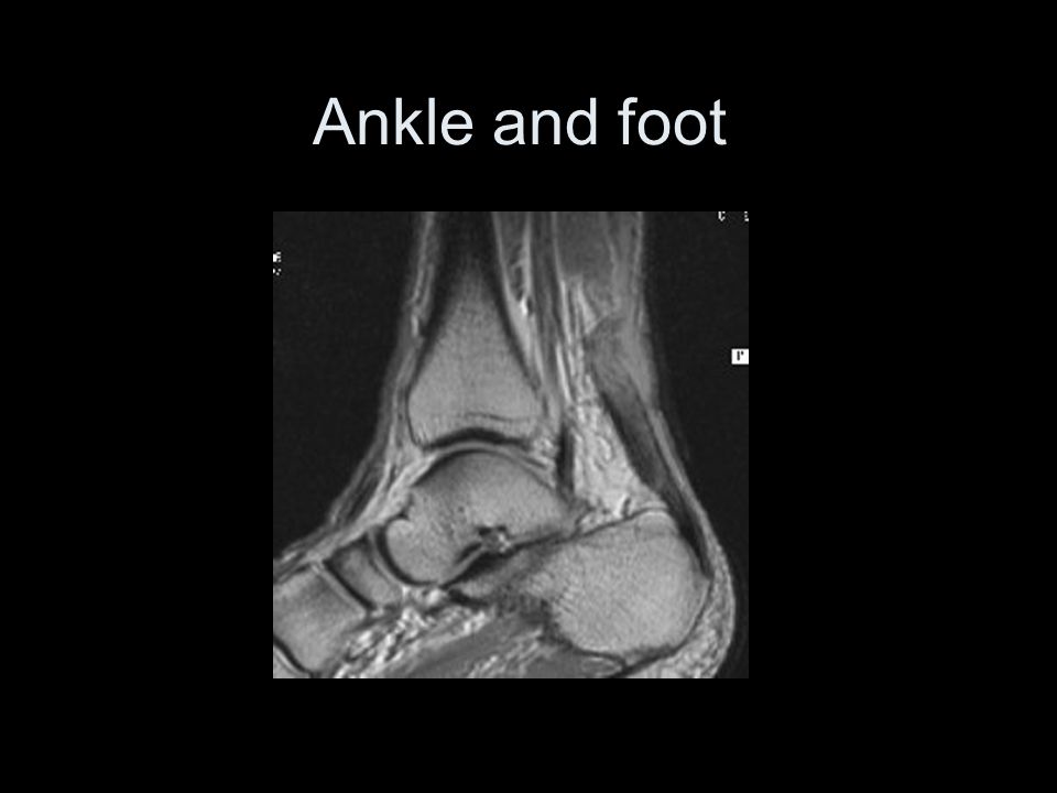 Ankle And Foot Saggital Slice Mri Ppt Video Online Download

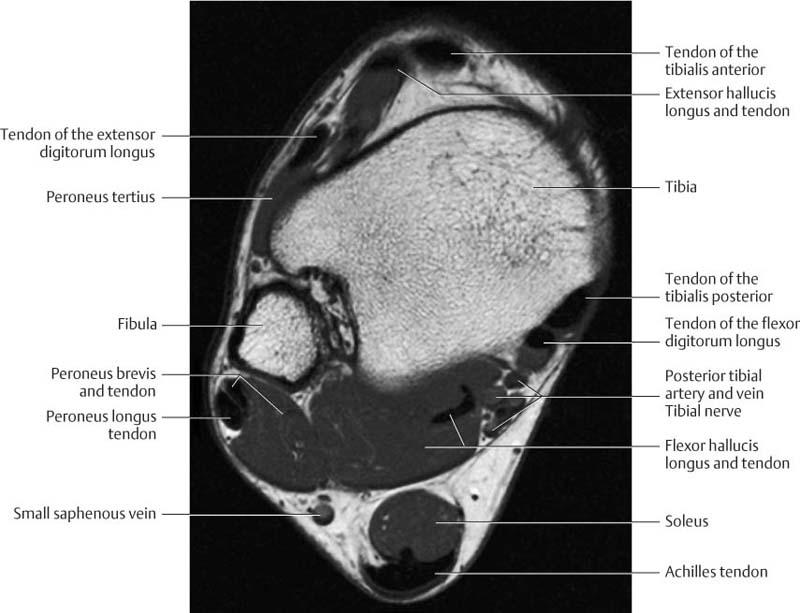
 Ankle Tendons Topographic Anatomy Radiology Case
 O Scan Esaote
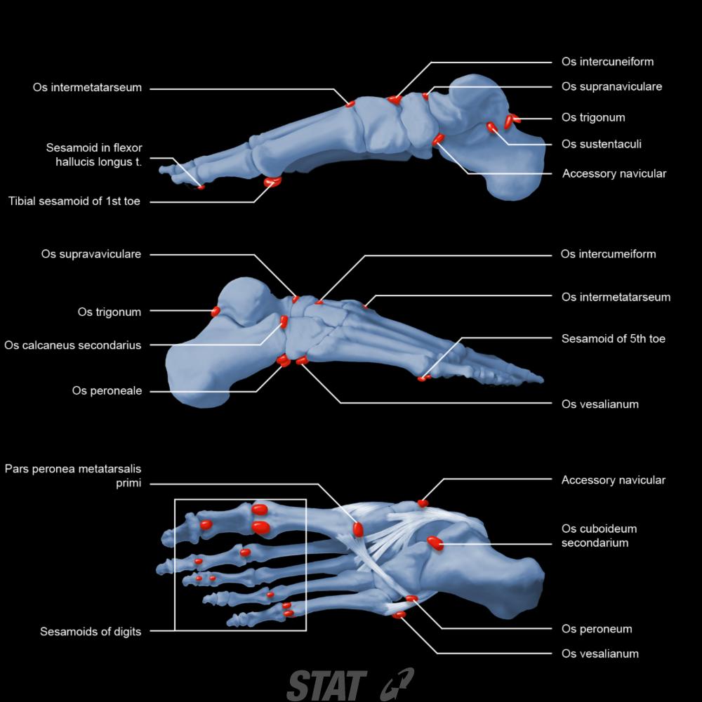
 Lower Extremity Os Foot Ankle Orthobullets
 Anatomy Of The Foot And Ankle Mri
Stanford Msk Mri Atlas C 2019
 The Knee Mri Atlas Of Anatomy In Medical Imagery
 An Mri Analysis Of The Pelvis To Determine The Ideal Method
 Ankle And Foot Radiology Key
 Mri Anatomy Of Ankle Radiology Case Radiopaedia Org
How To Read Knee Mri Of Normal Knee Anatomy Of The Knee Colorado Knee Specialist
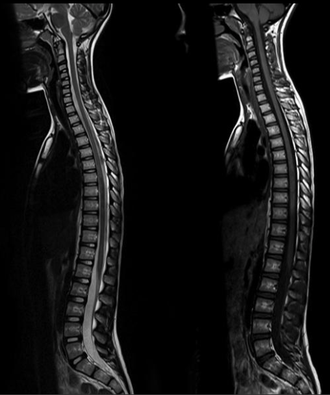 Normal Images Of Spine Joshua Li Md Phd


 The Radiology Assistant Ankle Mri Examination
Magnetic Resonance Imaging Of The Forefoot A Concise
 Anatomy Of The Foot And Ankle Mri
Belum ada Komentar untuk "Foot Anatomy Mri"
Posting Komentar