Anatomy Of Knee Meniscus
We hope this picture meniscus anatomy diagram can help you study and research. Two wedge shaped pieces of cartilage act as shock absorbers between your thighbone and shinbone.
 10093 01xv2 Left Knee Normal Anatomy Anatomy Exhibits
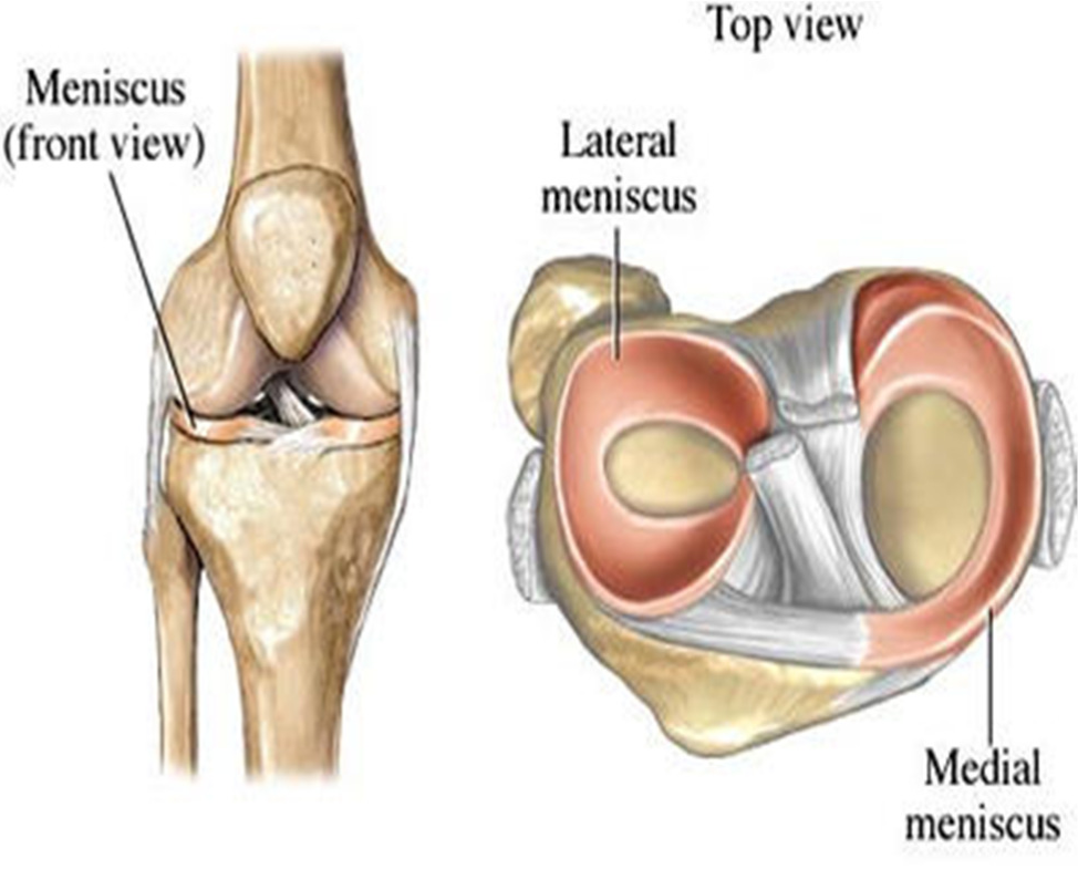
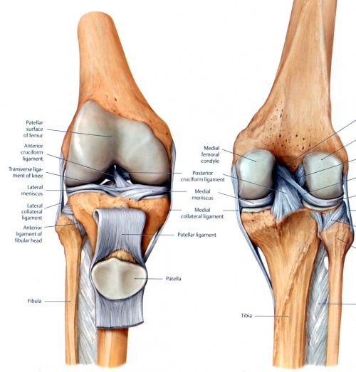
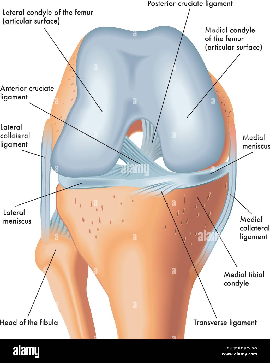
The knee joint contains the meniscus structure comprised of both a medial and a lateral component situated between the corresponding femoral condyle and tibial plateau figure 1.
Anatomy of knee meniscus. The knee meniscus is a special layer of extra cartilage that lines the knee joint. Pain especially when twisting or rotating your knee. Feeling as though your knee is locked in place when you try to move it.
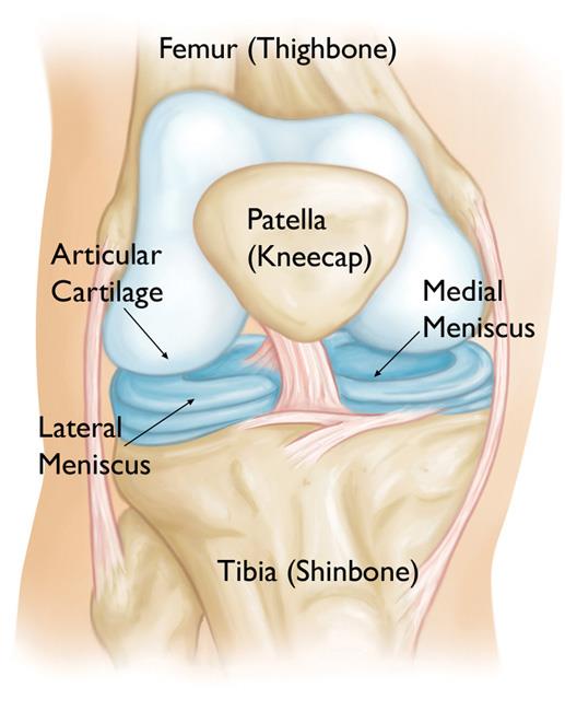
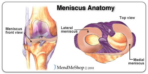
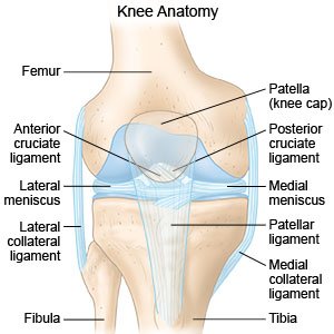
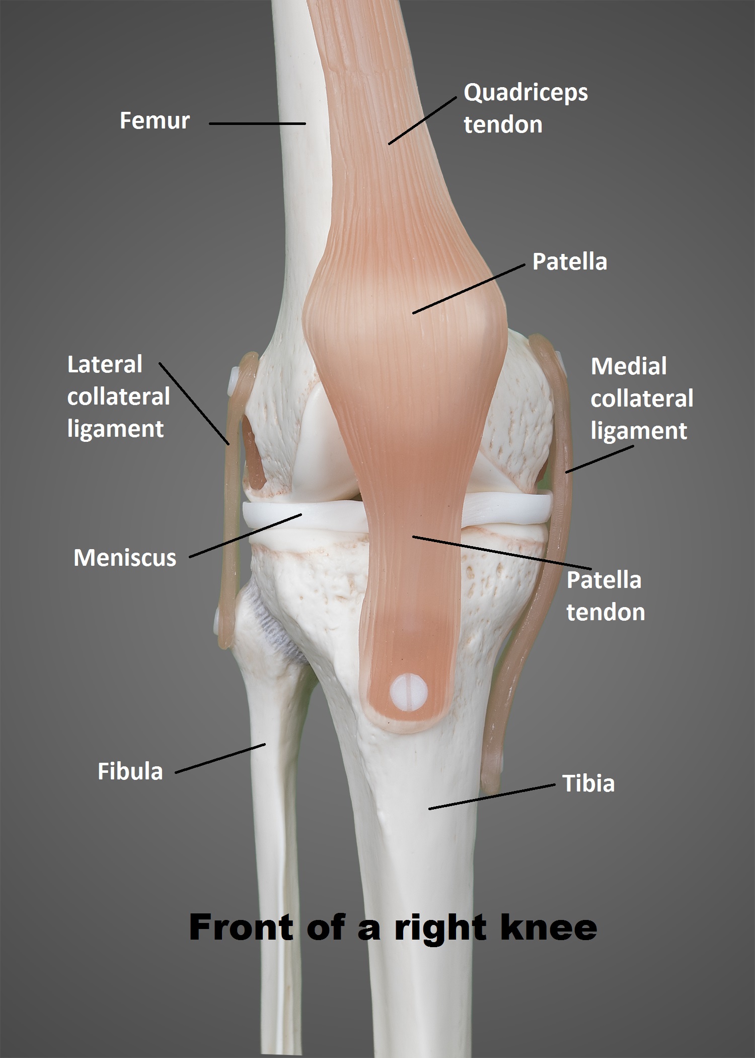
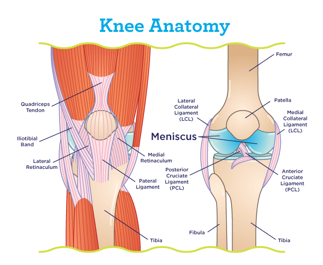
For more anatomy content please follow us and visit our website. Meniscus anatomy the menisci of the knee are two pads of fibrocartilaginous tissue which serve to disperse friction in the knee joint between the lower leg tibia and the thigh femur. Your thighbone femur shinbone tibia and kneecap patella.
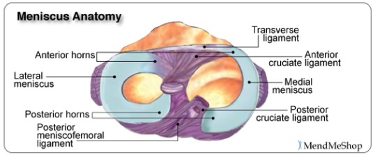
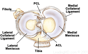
In cross section they have a triangular bow tie shape being thicker peripherally and thinning to a free edge centrally. The knee joins the thigh bone femur to the shin bone tibia. Each meniscus has a differing shape size and attachments.
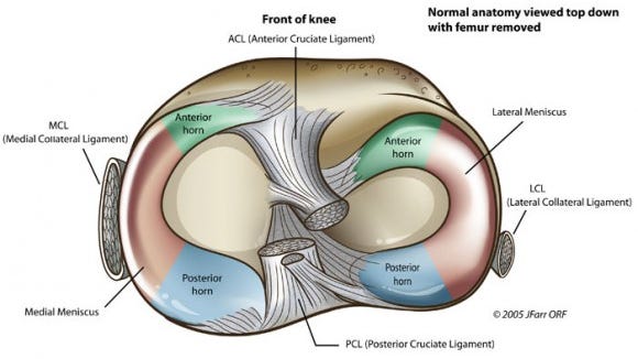
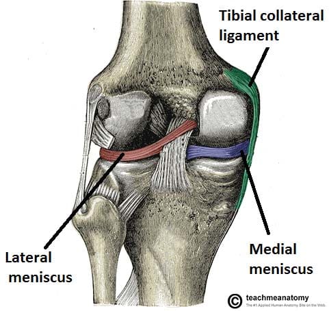
The band goes around the knee joint in a crescent shaped path and is located between the medial condyles of the shin and the femur or thighbone. The menisci are described as having a central body with anterior and posterior horns. It provides a smooth surface over the bones.
They are tough and rubbery to help cushion the joint and keep it stable. In most of our joints including the knee there is a layer of articular cartilage which is made of collagen and chondroitin. We think this is the most useful anatomy picture that you need.
The medial condyles are areas of these bones located on the inner sides of the knees. They are attached to the small depressions fossae. The smaller bone that runs alongside the tibia fibula and the kneecap patella are the other bones that make the knee joint.
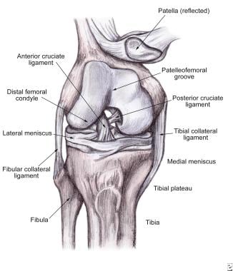
Its job is to cushion the joint and transfer forces between the tibia and femur bones. If youve torn your meniscus you might have the following signs and symptoms in your knee. Three bones meet to form your knee joint.
They are concave on the top and flat on the bottom articulating with the tibia. These are called meniscus. The knee is one of the largest and most complex joints in the body.
The medial meniscus is the central band of cartilage attached to the tibia or shinbone. Tendons connect the knee bones to the leg muscles that move the knee joint. Difficulty straightening your knee fully.
There are two knee menisci in each joint. Each is a glossy white complex tissue comprised of cells specialized extracellular matrix ecm molecules and region specific innervation and vascularization.
 Meniscal Tears Brisbane Knee And Shoulder Clinic Dr
 The Injury Zone Basic Anatomy And Function Of The Meniscus
 Meniscus Knee Sports Orthobullets
 Coronary Ligament Of The Knee Wikipedia
:max_bytes(150000):strip_icc()/vector-illustration-of-a-meniscus-tear-and-surgery-871162428-03ac23d73f854954a8082f2ae3ce9219.jpg) Meniscus Vs Cartilage Tear Of The Knee
Meniscus Vs Cartilage Tear Of The Knee
 Soft Tissue Knee Injury Practice Essentials Background
 Knee Physiopedia

 Posterior Cruciate Ligament An Overview Sciencedirect Topics
 Discoid Meniscus Orthoinfo Aaos
 Acl Solutions Acl Knee Anatomy And Diagram Images
 Acl Solutions Acl Knee Anatomy And Diagram Images
 Knee Sprain How To Treat A Sprained Knee
 The Knee Ut Health San Antonio
 Meniscus Injuries
 Injuries Of The Meniscus Of The Knee Sports Medicine
 Lateral Meniscus An Overview Sciencedirect Topics
 How Long Does It Take To Walk Or Work After Meniscus Repair
 Lateral Meniscus Wikipedia

 Meniscus Knees Knee Legs Skeleton Leg Thigh Joints
 Pin On All About Knee Pain
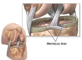
 Meniscus Tear
 The Knee Joint Articulations Movements Injuries
Belum ada Komentar untuk "Anatomy Of Knee Meniscus"
Posting Komentar