Anatomy Of Sinuses In Head
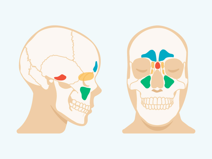
Humidify and filter the air while also producing mucus 4. The frontal sinuses lie behind the forehead above the eyes.
 Introduction To Biology Of The Ears Nose And Throat Ear
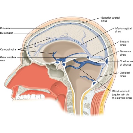
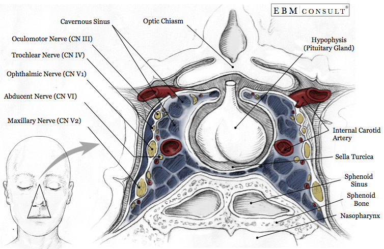
This sinus receives blood from the superior and inferior ophthalmic veins the middle superficial cerebral veins and from another dural venous sinus.
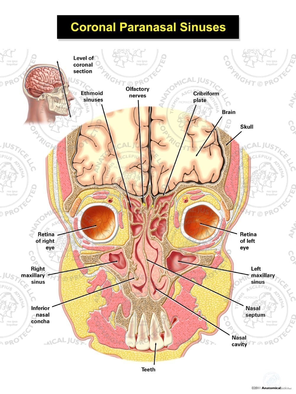
Anatomy of sinuses in head. The largest sinus cavities are about an inch across. The cavernous sinuses are a clinically important pair of dural sinuses. Sinuses definition sinuses are air filled spaces in the head that have several proposed functions.
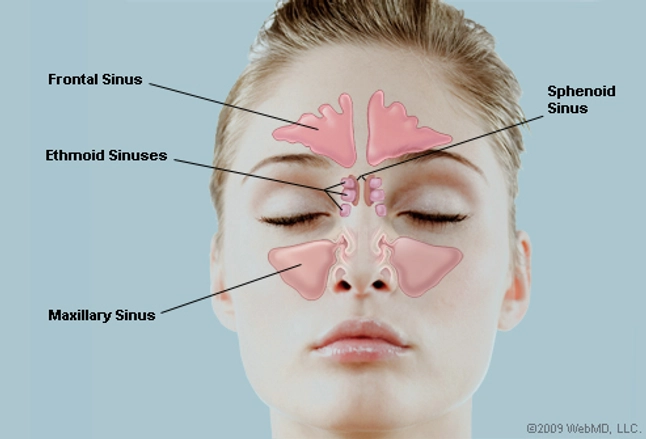
Play a role in vocal resonance. Your cheekbones hold your maxillary sinuses the largest. Most individuals have four paired cavities located in the cranial bone or skull.
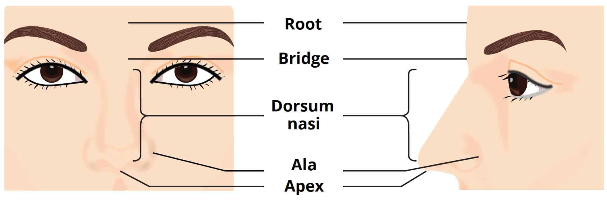
Anatomy of the sinuses. The sphenoid sinuses are located behind the ethmoid sinuses. This bone separates the eyes from the nose.
Lighten the skull 3. We think this is the most useful anatomy picture that you need. The sinuses themselves are separated from the eye and the nose by very thin bone or bony laminae.
Sinus anatomy in common usage sinus usually refers to the paranasal sinuses which are air cavities in the cranial bones especially those near the nose and connecting to it. There are four pairs of paranasal sinuses the frontal sinuses are located above the eyes in the forehead bone. When they arent moistening the air we breathe through our noses.
The maxillary sinuses are behind the cheeks. The sinuses are lined with mucus producing membranes that help guard against pathogens debris and pollutants. Between your eyes are your ethmoid sinuses.
Anatomy lecture 1 cranial cavity dural sinuses innervation of head neck. The sphenoid sinuses are near the base of the skull and can even spread as far as the occipital bone near the back of the head if they are very large. There are several sets of sinuses in the head.
The ethmoid sinuses also called ethmoid labyrinth are located between the eyes and the nose. Anatomy of the sinuses. Like the nasal cavity the sinuses are all lined with mucus.
The low center of your forehead is where your frontal sinuses are located. They are located next to the lateral aspect of the body of the sphenoid bone. They serve as shock absorbers in cases of head trauma 2.
The mucus secretions produced in the sinuses are continually being swept into the nose by the hair like structures called cilia on the surface of the respiratory membrane. The head contains 4 paired sinus cavities. For more anatomy content please follow us and visit our website.
The sphenoid sinuses are. The ethmoid sinuses lie under the inside corners of the eyes. The maxillary sinuses antra of highmore are located in the cheekbones under the eyes.
Others are much smaller. We hope this picture sinus anatomy in head and sinusitis can help you study and research. The sinuses are a connected system of hollow cavities in the skull.
 Paranasal Sinus An Overview Sciencedirect Topics
 Anatomy Of Sinuses Brain Sinus Anatomy Human Anatomy Diagram
 The Paranasal Sinuses Structure Function Teachmeanatomy
 Dural Venous Sinuses Radiology Reference Article
 Figure Nasal Cavity Frontal Sinus Superior
 The Nose And Sinuses Teachmeanatomy
 Cranial Nerve Anatomy Cranial Nerves Iowa Head And Neck
Chronic And Recurrent Sinusitis Ear Nose Throat
:background_color(FFFFFF):format(jpeg)/images/library/10270/Paranasal_Sinuses.png) Schwannoma Of The Nasal Cavity Clinical Case Diagnosis
Schwannoma Of The Nasal Cavity Clinical Case Diagnosis
 Chapter 23 Nasal Cavity The Big Picture Gross Anatomy
 Sinus Cavities In The Head Anatomy Diagram Pictures
 Nasal Sinus Cavity Anatomy Sinus Cavities Paranasal
 Sinusitis Allergy Asthma And Sinus Specialists City Allergy
 What Are The Sinuses Pictures Of Nasal Cavities
 Tri 2 Gross Anatomy Sinuses Diagram Quizlet
 Section Of Head Model Sagital Section Of Head
:watermark(/images/watermark_only.png,0,0,0):watermark(/images/logo_url.png,-10,-10,0):format(jpeg)/images/anatomy_term/sinus-sagittalis-superior/FHOctzOmsy0wGTVWVWmgzg_Superior_sagittal_sinus_01.png) Dural Venous Sinuses Anatomy Kenhub
Dural Venous Sinuses Anatomy Kenhub
 What Are Sinuses Anatomy Types Study Com
 Sinus Cancer Anatomy Headandneckcancerguide Org
 Ecr 2017 C 2117 Ct Anatomy Of Paranasal Sinuses Epos
 Sinus Cavities In The Head Anatomy Diagram Pictures
 Equine Sinus Disease A Hidden Danger Expert How To For


Belum ada Komentar untuk "Anatomy Of Sinuses In Head"
Posting Komentar