Anatomy Of Middle Ear
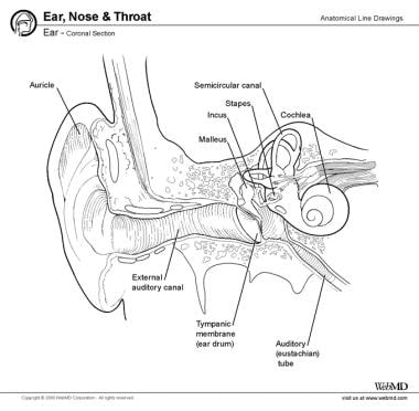
Auditory tube drains fluid from the. The ossicles directly couple sound energy from the ear drum to the oval window of the cochlea.
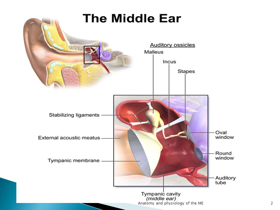 Anatomy And Physiology Of The Middle Ear Ppt Video Online
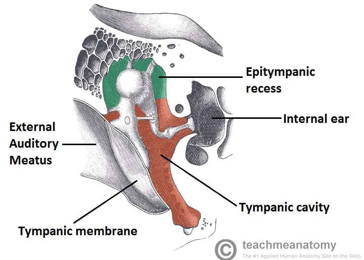
Middle ear anatomy tympanic cavity is an air filled cavity.
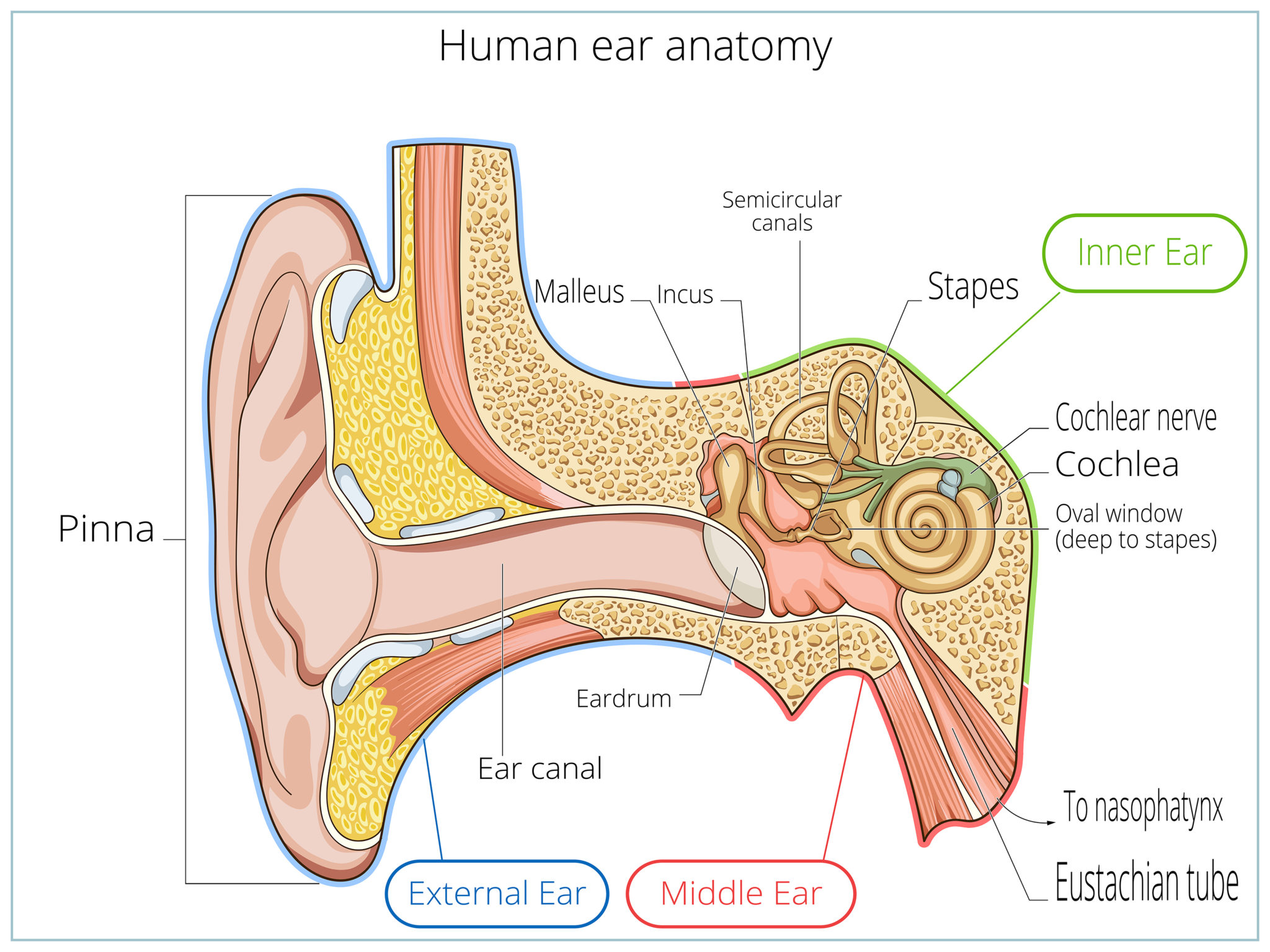
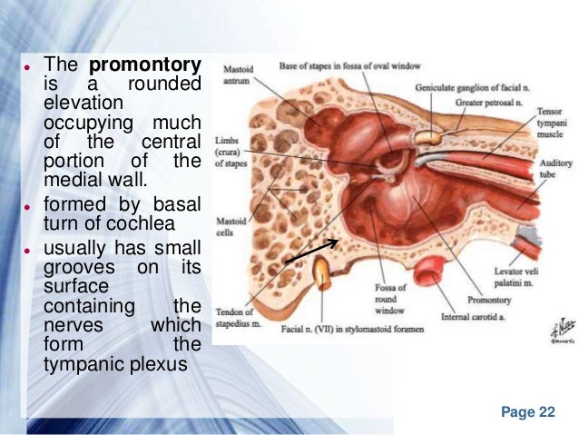
Anatomy of middle ear. Middle ear tympanic cavity consisting of. Sam webster 38861 views. The middle ear also known as the tympanic cavity or the tympanum is a pneumatized air filled region of the temporal bone that lies just medial to the tympanic membrane ear drum and lateral to the promontory caused by the turns of the cochlea of the ear.
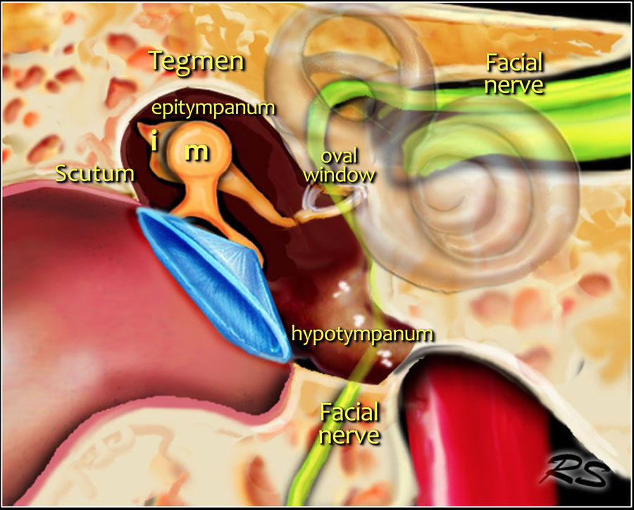
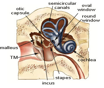
Oval window connects the middle ear with the inner ear. The bones are called. The inner ear includes.
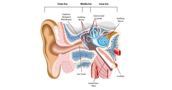
The chambers are full of fluid which vibrates when sound comes in and causes the small hairs which line the membrane to vibrate and send electrical impulses to the brain. It is also membrane lined interplanetary cavity situated between the ear canal and the eustachian tube cochlea and auditory nerve. The cochlea which is the hearing portion and the semicircular canals is the balance portion.
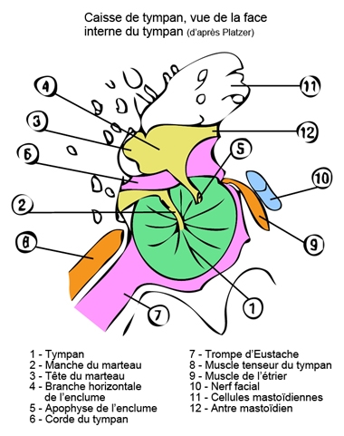
Malleus incus and stapes. The eardrum splits this cavity from the ear canal. The middle ear contains three tiny bones known as the ossicles.
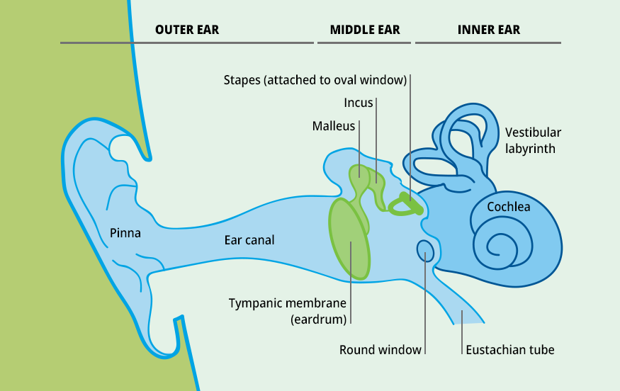
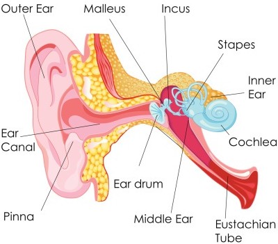
The outer ear is called the pinna and is made of ridged cartilage covered by skin. The eardrum acts as a natural boundary between the middle ear and the ear canal. Three small bones that are connected and transmit the sound waves to the inner ear.
The eardrum separates this space from the ear canal. Transforms sound into signals that get sent to the brain. A canal that links the middle ear with the back of the nose.
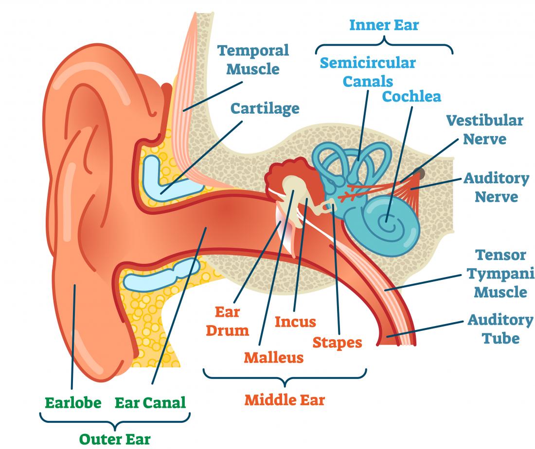
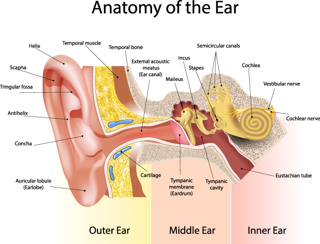
Ear anatomy inner ear. The ossicles were given their latin names for their distinctive shapes. The ear has external middle and inner portions.
Also known as the tympanic cavity the middle ear is an air filled membrane lined space located between the ear canal and the eustachian tube cochlea and auditory nerve. Semicircular ducts filled with fluid. The area is pressurized.
Sound funnels through the pinna into the external auditory. The cochlea is shaped like a snail and is divided into two chambers by a membrane. Middle ear tympanic cavity anatomy duration.
Cochlea spiral shaped organ of hearing. They are also referred to as the hammer anvil and stirrup respectively. Attached to cochlea and nerves.
The eustachian tube helps to equalize the pressure in.
 Patient And Parent Education
 Anatomy Of The Ear
 Ear Muscle Anatomy
 Hearing Loss Education Facts And Causes How We Hear
 Middle Ear Wikipedia
 Middle Ear Anatomy Www Medicoapps Org
 Ear Anatomy Healthlink Bc
 Ears
Ears
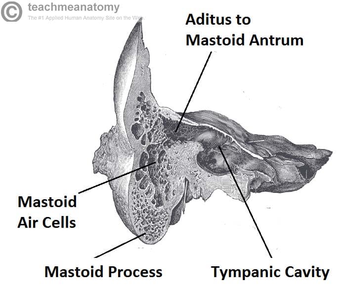 The Middle Ear Parts Bones Muscles Teachmeanatomy
 The Radiology Assistant Temporal Bone Anatomy 2 0
 Middle Ear Anatomy At Grand Valley State University Studyblue
 Middle Ear Cavity Middle Ear Middle Ear Anatomy Ear Anatomy
 Oval Window Wikipedia
 Middle Ear Anatomy
 Middle Ear Anatomy Images And Video Britannica Com
:max_bytes(150000):strip_icc()/GettyImages-506836745-595d4dc05f9b58843f509ed2.jpg) Patulous Eustachian Tube Symptoms Causes And Treament
Patulous Eustachian Tube Symptoms Causes And Treament
 Ears And Hearing How Do They Work
 The Human Ear Consists Of Three Parts The Outer Ear Middle Ear
 The Anatomy Of The Human Ear The Middle Ear Health Life
 Anatomy Of The Middle Ear A Schematic Of A Sauropsid
 Anatomy Of Middle Ear With Clinical Correlation Epomedicine
 Ear Anatomy
 Bountiful Layton Ear Tube Surgery For Middle Ear Infection
 Ear Anatomy Overview Embryology Gross Anatomy

 Unit One Normal Anatomy
 Middle Ear Conditions Anatomical Chart
 Anatomy Of Middle Ear With Clinical Correlation Epomedicine
 External And Middle Ear Anatomy Diagram Quizlet
 The Middle Ear Parts Bones Muscles Teachmeanatomy
Belum ada Komentar untuk "Anatomy Of Middle Ear"
Posting Komentar