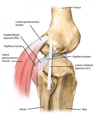
Lcl Anatomy
Injuries to the lcl and posterior lateral corner result from a rotational force across. Injury to the lateral collateral ligament lcl also referred to as fibular collateral ligament.
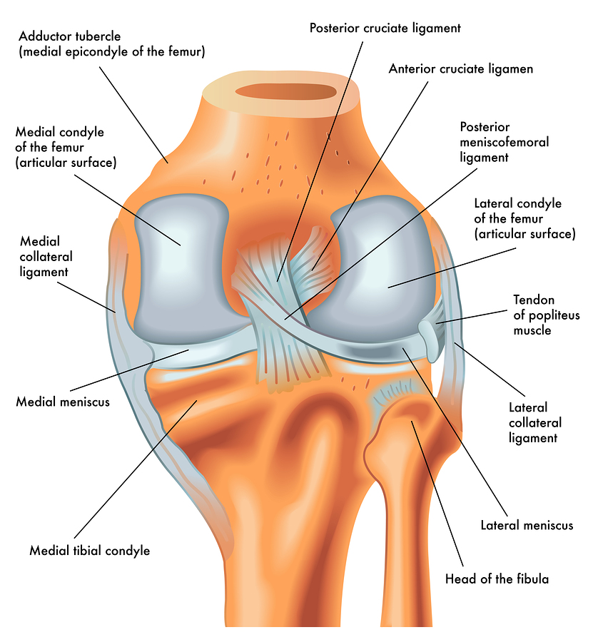
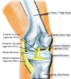
 Anatomy Of The Knee
Acl anterior cruciate ligament ant anterior itt iliotibial tract lcl lateral collateral ligament mcl medial collateral ligament pcl posterior cruciate ligament post posterior pt popliteus muscle and tendon sm semimembranosus tendon.

Lcl anatomy. Gross anatomy it originates from the lateral femoral epicondyle and has an oblique course is join. When the lcl does rip it will need serious treatment and reconstruction. The popliteal tendon pt is considered to be a dynamic stabilizer and the lateral collateral ligament lcl the fabellofibular ligament ffl the popliteofibular ligament pfl.
The lateral collateral ligament lcl is a one of the four major ligaments in the knee. A careful eye is needed to diagnose a posterolateral and lateral. The lcl runs along the outside of the knee joint from the outside of the bottom of the thighbone femur to the top of the lower leg bone fibula.
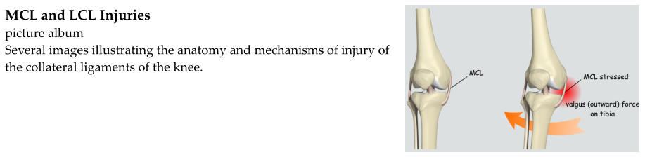
The collateral ligaments control the sideways motion of your knee and brace it against unusual movement. 41 anatomy and normal mri appearance. Lateral collateral ligament lcl two of the knee ligaments are collateral or toward the side of the knee joint.
It is one of 4 critical ligaments involved in stabilizing the knee joint. The fibular or lateral collateral ligament lcl is a cord like band and acts as the primary varus stabilizer of the knee. The lateral collateral ligament or lcl faces outward.
The lateral or outside collateral ligament lcl connects the femur to the smaller bone in the lower leg fibula. Ligaments are thick strong bands of tissue that connect bone to bone. 7 16 of all knee ligament injuries when combined with lateral ligamentous complex injuries.
The lateral collateral ligament lcl is the ligament located in the knee joint. The fibular collateral ligament long external lateral ligament or lateral collateral ligament lcl is a ligament located on the lateral outer side of the knee and thus belongs to the extrinsic knee ligaments and posterolateral corner of the knee. The lcl may tear after a hard hit to the outside of the knee but other ligaments are more likely to tear.
The posterolateral corner limits posterior translation varus angulation and excessive external rotation. Epidemiology demographics incidence isolated injury extremely rare. The lateral fibular collateral ligament is a cord like ligament on the lateral aspect of the knee and forms part of the posterolateral corner.
 Lateral Collateral Ligament
 Mcl Knee Injuries Knee Sports Orthobullets
 Acl Mcl Lcl Diagram Reading Industrial Wiring Diagrams
 Acl Mcl Lcl Diagram Reading Industrial Wiring Diagrams
 Lcl Injury Active Care Physiotherapy Clinic
 Jay S Physio Lcl What You Know About Lateral Collateral
 Knee Pain And Injuries Injury Center Of Houston
 Lcl Injury
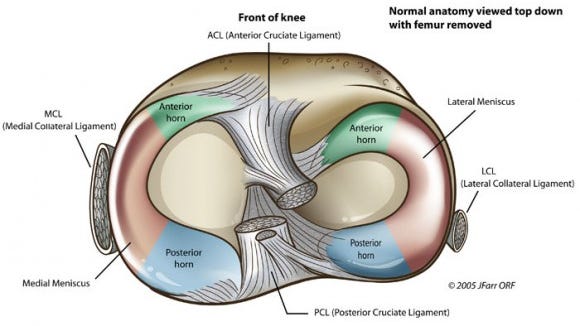 The Injury Zone Basic Anatomy And Function Of The Meniscus
 Dial Test Physiopedia
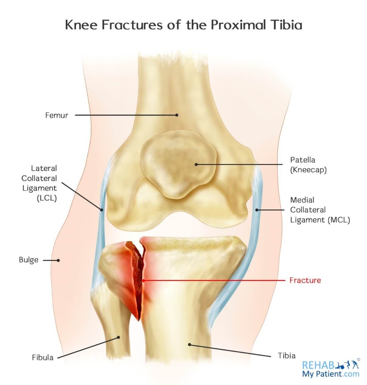 Knee Fractures Of The Proximal Tibia Rehab My Patient
 Collateral Ligament Injuries Orthoinfo Aaos
 Posterolateral Corner Injury Knee Sports Orthobullets
 Posterolateral Corner Injuries Epidemiology Anatomy
Knee Sprain Information And Treatment For Ligament Knee Inuries
 Lcl Plc Reconstruction Gavin Mchugh
 Elbow Anatomy Biomechanics Shoulder Elbow Orthobullets
 Lcl Injury Of The Knee Knee Sports Orthobullets
 The Effect Of Prolonged Knee Extension Immobilization On
 Lcl Plc Reconstruction Gavin Mchugh
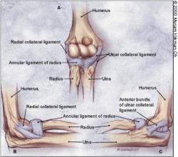 Elbow Ligamentous Injuries Physiopedia
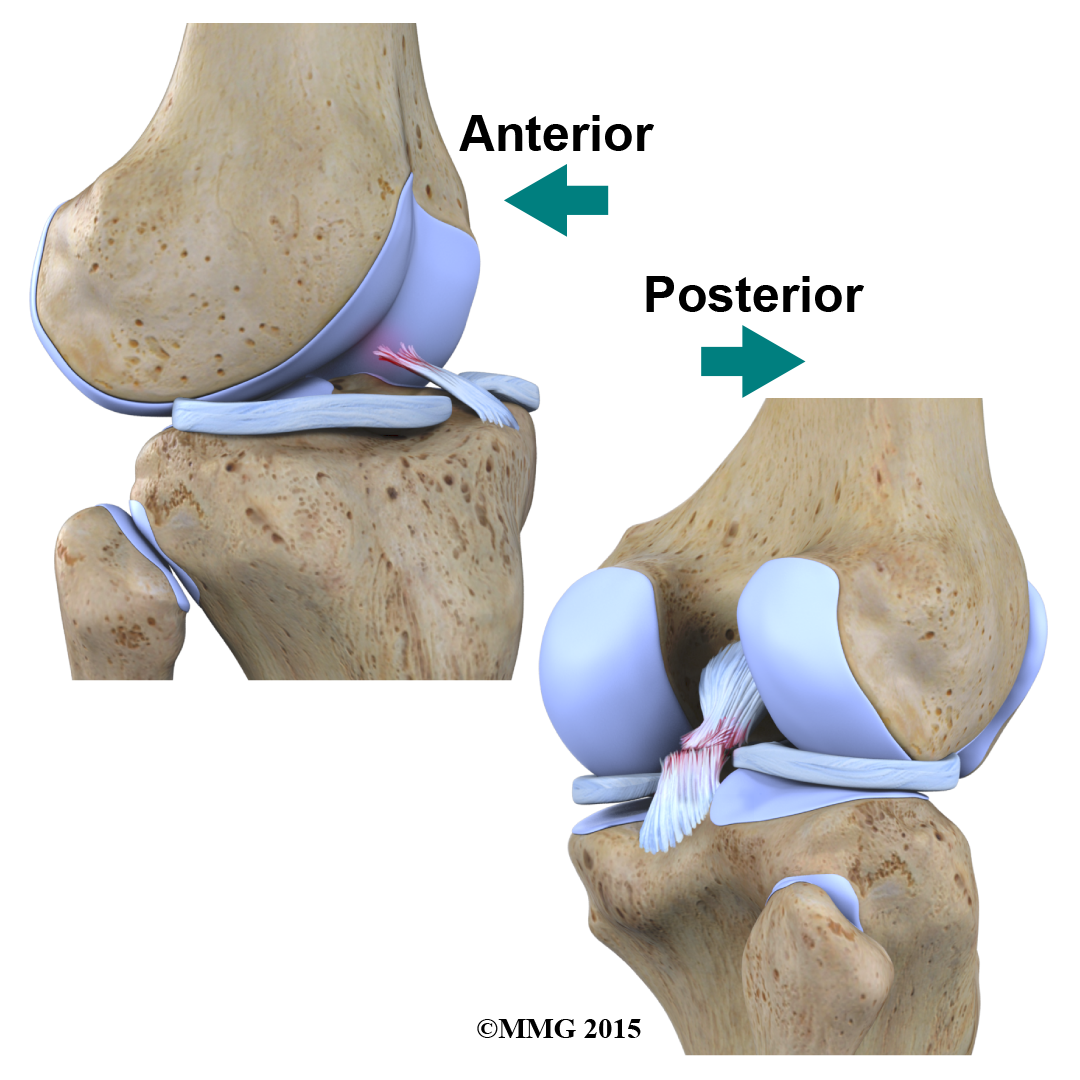 Physical Therapy In Conway For Knee Anatomy
 Lateral Meniscus Wikipedia
 Posterolateral Corner Injuries Epidemiology Anatomy
 Lateral Collateral Ligament And The Nfl Football Player
Ligament Injuries To The Knee Johns Hopkins Medicine
 Collateral Ligament Injuries Of The Knee
 Lcl Injury Of The Knee Knee Sports Orthobullets

Belum ada Komentar untuk "Lcl Anatomy"
Posting Komentar