Scalene Anatomy
The name refers to the shape that is formed when the three scalene muscles come together on each side of the neck to form a scalene triangle. For that reason they are also considered as accessory muscles of inspiration.

The anterior and middle scalene muscles lift the first rib and bend the neck to the same side.

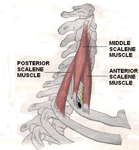
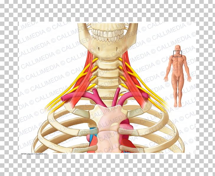
Scalene anatomy. The scalene muscles are involved lifting the first two ribs in a forced inspiratory act as secondary respiratory muscles. The scalene muscles elevate the ribs and therefore the thorax. The posterior scalene lifts the second rib and tilts the neck to the same side.
There is an anterior scalene scalenus anterior a medial scalene scalenus medius and a posterior scalene scalenus posterior. To strength scalene sit in a comfortable chair. The scalene muscles are a major contributor to thoracic outlet syndrome thoracic outlet syndrome tos happens when the nerves andor blood vessels running from the neck down into the arm are compressed in an area between the base of the neck and armpit.
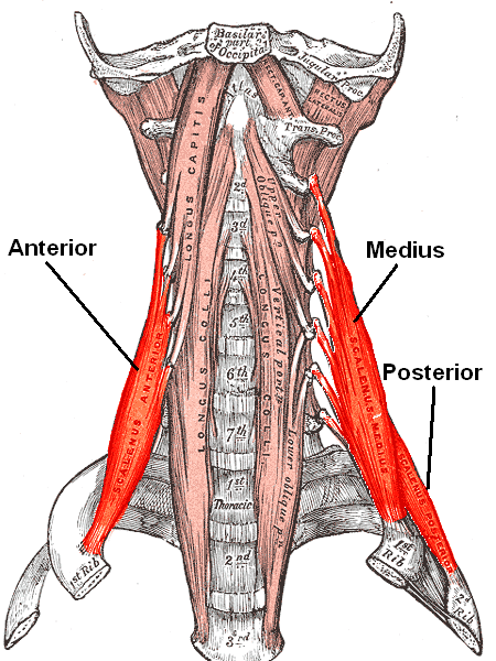
They are innervated by the fourth fifth and sixth cervical spinal nerves. Hold the ends of the band. The scalene muscles are a group of three pairs of muscles in the lateral neck namely the anterior scalene middle scalene and posterior scalene.
The scalene muscles are three paired muscles of the neck located in the front on either side of the throat just lateral to the sternocleidomastoid. In reality the scalene muscles are always electrically active even for not necessarily forced breaths. Place the palm of your right hand on the right side of your head.
A unilateral contraction bends the cervical spine to the side. The muscles are named from ancient greek σκαληνός meaning uneven. Ipsilateral contraction causes ipsilateral lateral flexion of the neck.
Anterior and medial scalene elevate first rib and flexes neck to same side. Scalene muscles have three main functions. The name scalene is related to the greek word skalenos which was used to refer to a triangle of unequal sides.
Elevation of the first rib. Originates from the posterior tubercles of the transverse processes of c2 c7 and attaches to the scalene tubercle of the first rib. Posterior scalene elevates second rib and flexes neck to same side.
This hand acts as a stabilizer in the exercise. Another exercise to strength scalene. The scalene muscles when not engaged as respiratory muscles are very active in the supine position and with the arm raised.
Sit or stand up straight with a resistance band looped around your head.
 Scalene Physiopedia
![]() Scalene Muscles Wikiwand
Scalene Muscles Wikiwand
 Sternohyoid Muscle Sternothyroid Muscle And Scalene
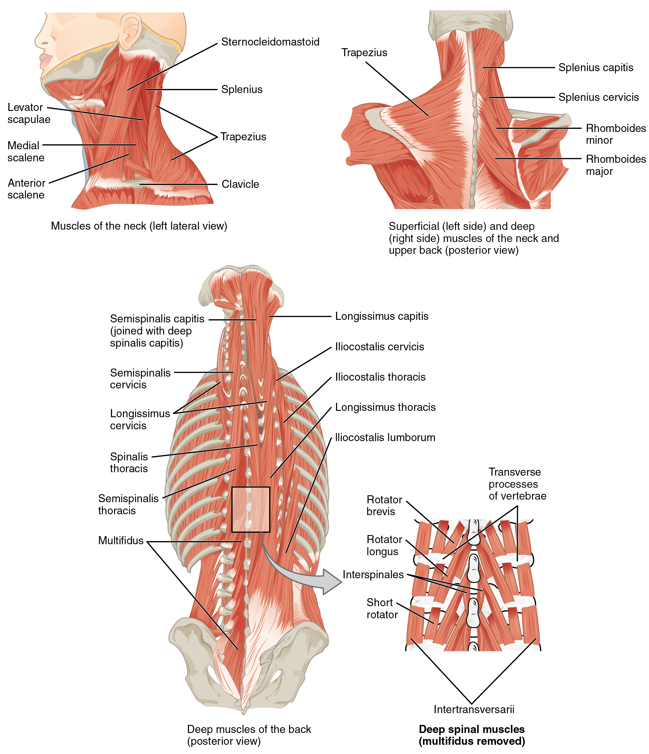 11 3 Axial Muscles Of The Head Neck And Back Anatomy And
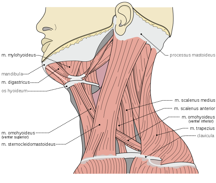 The Ventral Neck Muscles Lecturio Online Medical Library
 Thoracic Outlet Syndrome Tos Msk Medbullets Step 1
 Counting Scalenes Because Functional Anatomy Says So
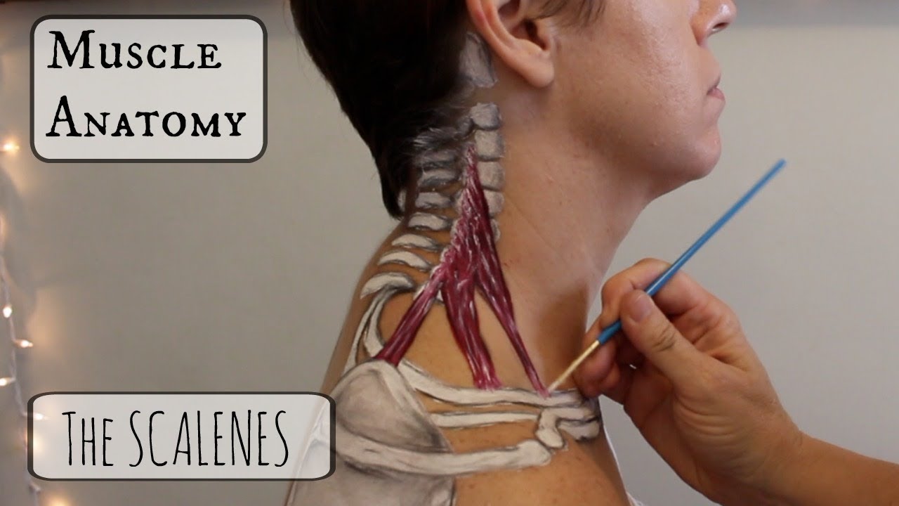 Muscle Anatomy The Scalenes
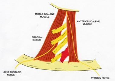
 Brachial Plexus Part 1 Anatomical Relations Sketchy Medicine
 Nerve Cervicobrachial Neuralgia Scalene Muscles Brachial
 Sports Physiotherapist On Twitter Thoracic Outlet Syndrome
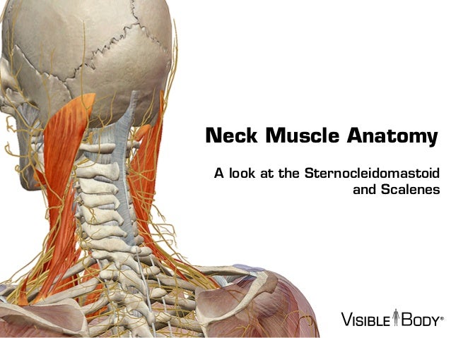 Visible Body Sternocleidomastoid And The Scalene Muscles
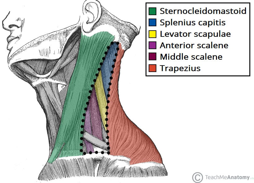 Posterior Triangle Of The Neck Subdivisions Teachmeanatomy
 Scalene Muscles An Overview Sciencedirect Topics
 Sternocleidomastoid And Scalenes Muscles Of The Neck
 Stock Illustration

 Long Thoracic Nerve Injury The Shortest Route To Recovery
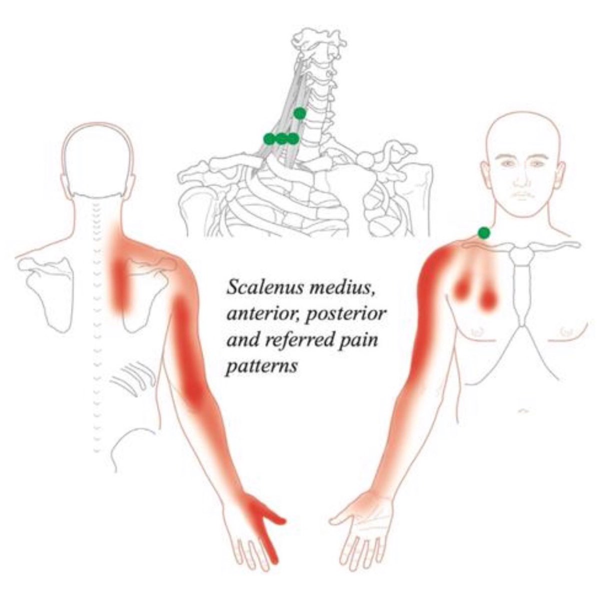
 Scalene Trigger Points And Referred Pain Patterns
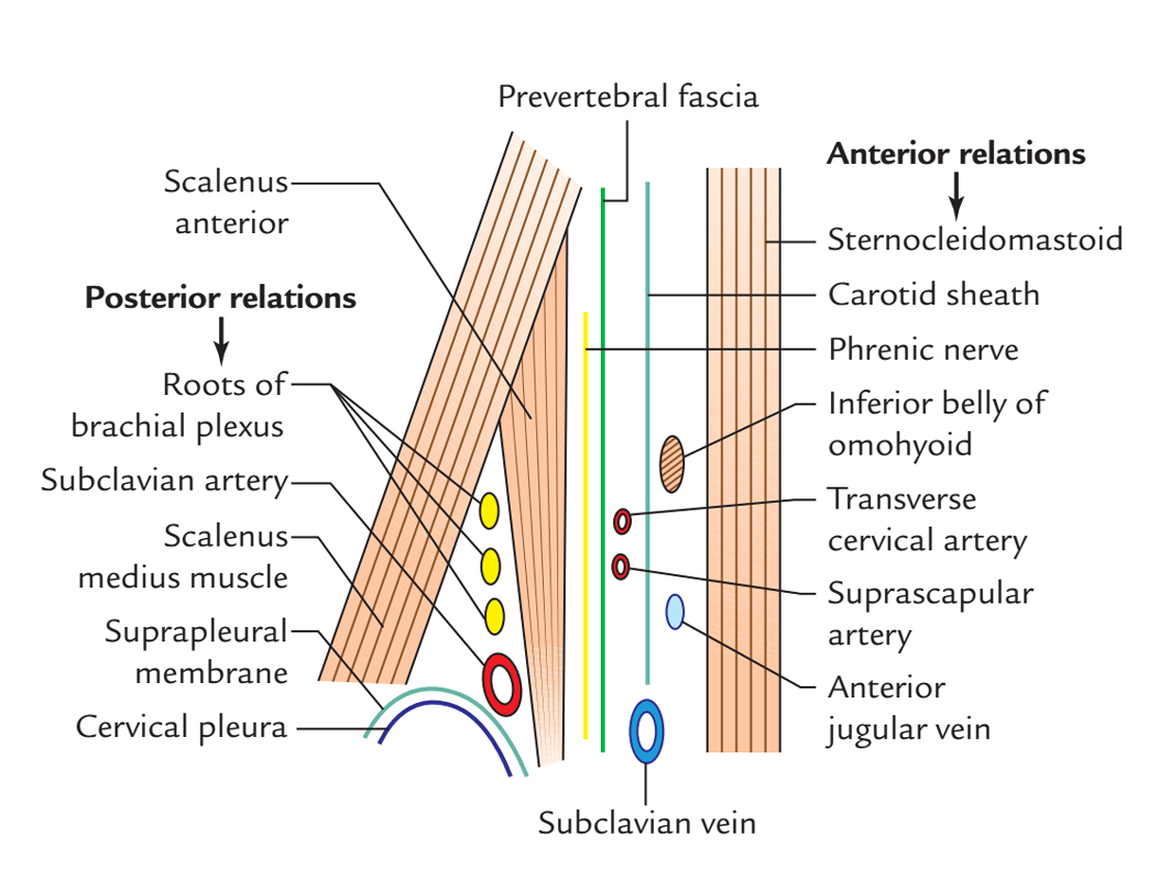
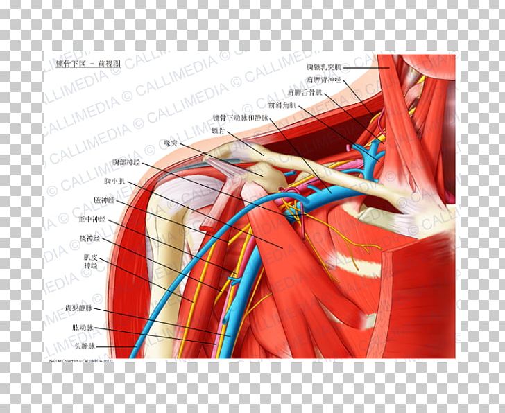 Infraclavicular Fossa Anatomy Subclavian Artery Scalene
 Neck Muscles Scalene Muscle And Digastric Muscle Anatomy
 Thoracic Outlet Obstruction Background Anatomy
 Scalene Muscles Functional Anatomyintegrative Works
 Scalene Muscles Wikipedia
 Anterior Scalene Muscle Scalenus Anterior Earth S Lab
 Ultrasound Guided Interscalene Brachial Plexus Block Nysora
 Scalene Anatomy Orthobullets
The Scalene Anterior Stock Illustration Illustration Of
Belum ada Komentar untuk "Scalene Anatomy"
Posting Komentar