Collateral Anatomy
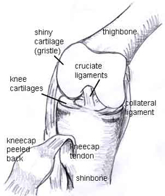
In anatomy a collateral is a subordinate or accessory part. Two c shaped pieces of cartilage called the medial and lateral menisci act as shock absorbers between the.
 Understanding The Medial Ulnar Collateral Ligament Of The
When stress is applied this ligament aids control in transferring the joint through a normal range of movement.

Collateral anatomy. However several of their branches can become important collateral pathways if occlusion occurs in the internal carotid or vertebral arteries. The mcl also prevents an anterior movement of the tibia and hyperextension. Annular ligament from.
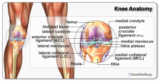
The collateral ligaments medial mcl and lateral lcl are found on the sides of your knee. A collateral is also a side branch as of a blood vessel or nerve. The medial and lateral collateral ligaments prevent the femur from sliding side to side.
The axon terminals send signals to the next nerve cell and so forth. A side branch of a nerve axon or blood vessel. The dendrites accept the signal received from the other nerve cells and the axon carried signals to the axon terminals.
The branches of the external carotid artery are the ascending pharyngeal the superior thyroid the lingual the external maxillary the occipital the facial the posterior auricular. The medial collateral ligament is recognised as being a primary static stabiliser of the knee and assists in passively stabilising the joint. Indirect subsidiary or accessory to the main thing.
The lateral radial collateral ligament lclrcl complex is a major lateral stabilizer of the elbow joint and resists varus stress. The structures that are considered static stabilizers of the medial knee are the superficial mcl the deep mcl and the posterior oblique ligament. A collateral is also a side branch as of a blood vessel or nerve.
These are often contact injuries but not always. Learn about financial products learn about real estate products. Gross anatomy the lcl is a y shaped ligamentous complex composed of three parts 1 2.
Collateral analytics ca develops real estate analytic products and tools to support financial institutions institutional and retail investors as well as property capital market activities. The superficial mcl is the primary static medial stabilizer of the knee situated in the second layer according to warren and marshalls three layer concept. The axon collateral can be a part of feedback mechanism which creates a connection with nearby inhibitory neurons and thus they can be involved in regulation of the neuron over excitation.
Injuries to the collateral ligaments are usually caused by a force that pushes the knee sideways.
 Fibular Collateral Ligament Injury Joint Anatomy Vector
 Gonadal Veins Normal Computed Tomography Anatomy And
 Human Anatomy And Physiology Diagrams Ulnar Collateral
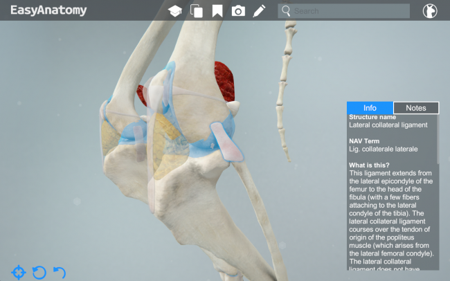 Canine Lateral Collateral Ligament Easyanatomy Easyanatomy
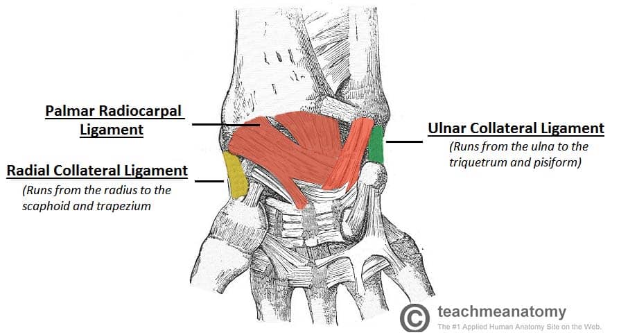 The Wrist Joint Teachmeanatomy
 Medial Collateral Ligament Mcl Injuries Thermoskin
 The Medial Collateral Ligament Part 1 Anatomy
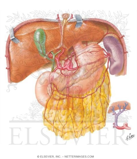 Arterial Variations And Collateral Supply Of Liver And
 Knee Anatomy Focus Pocus

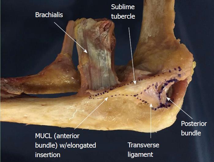
 Anatomy Of The Elbow Elbow Anatomy
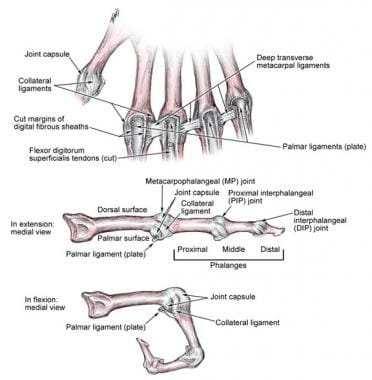 Metacarpophalangeal And Interphalangeal Ligament Anatomy
 Physical Therapy In Dothan For Knee Collateral Ligament Injury
 Introduction To The Collateral Ligaments Kneeguru
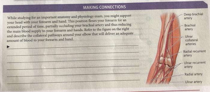 Solved Making Connections While Studying For An Important
Collateral Anatomy Exhibits


 File Anatomy Of The Ulnar Collateral Ligament In The
 Ulnar Collateral Ligament Anatomy Download Scientific Diagram
 Collateral Ligament An Overview Sciencedirect Topics
Collateral Ligament Injuries Orthoinfo Aaos
Belum ada Komentar untuk "Collateral Anatomy"
Posting Komentar