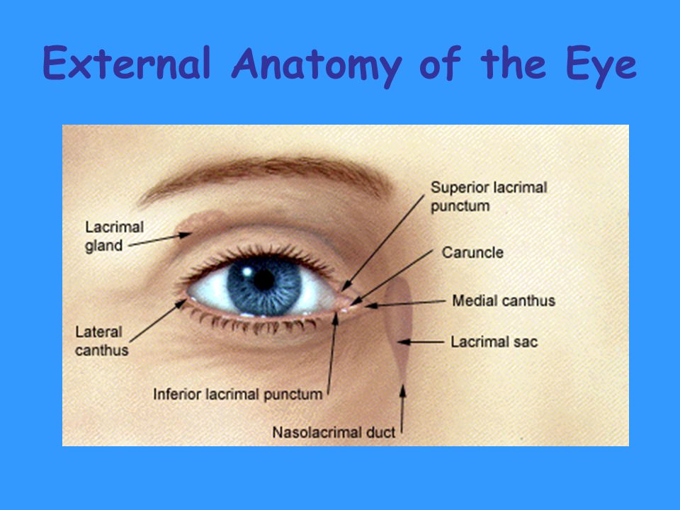
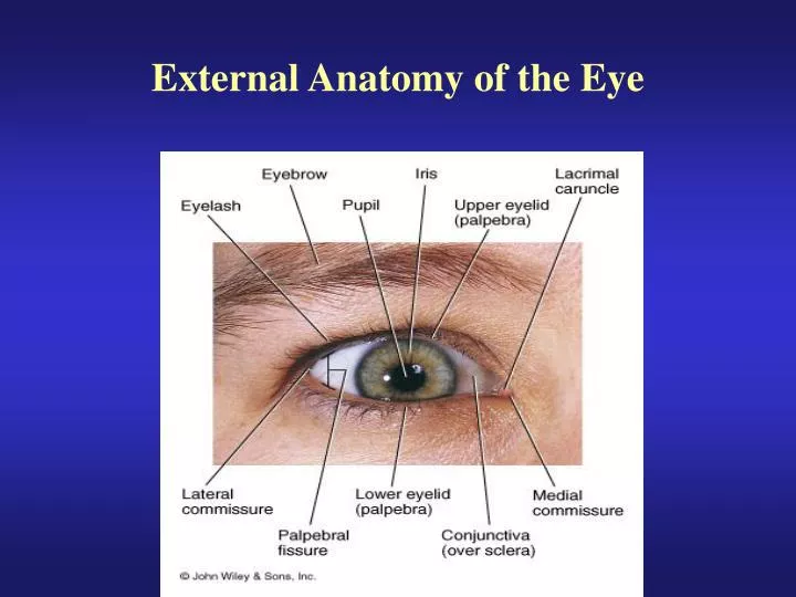
External Anatomy Of The Eye
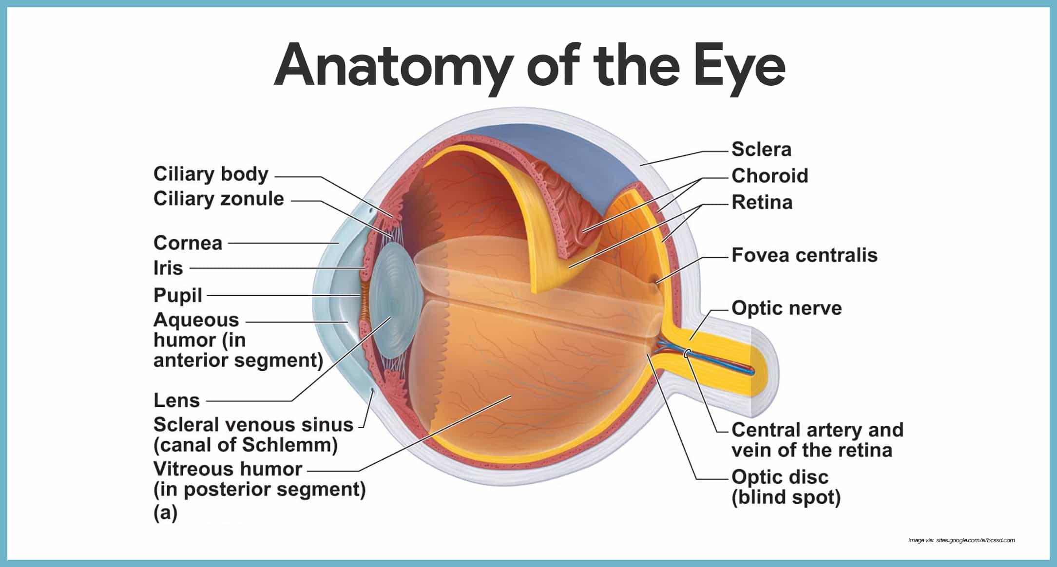
The structure of the human eye is made of three layers. Blinking on the other hand which is closing and opening the eye rapidly spreads tears across and and removes irritants from the cornea and conjuctiva.
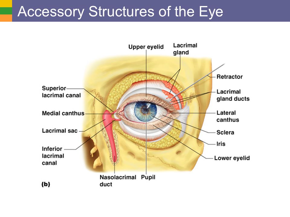
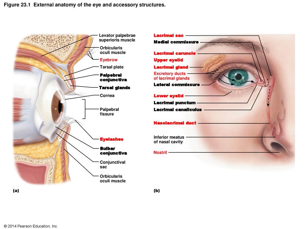 Figure 23 1 External Anatomy Of The Eye And Accessory
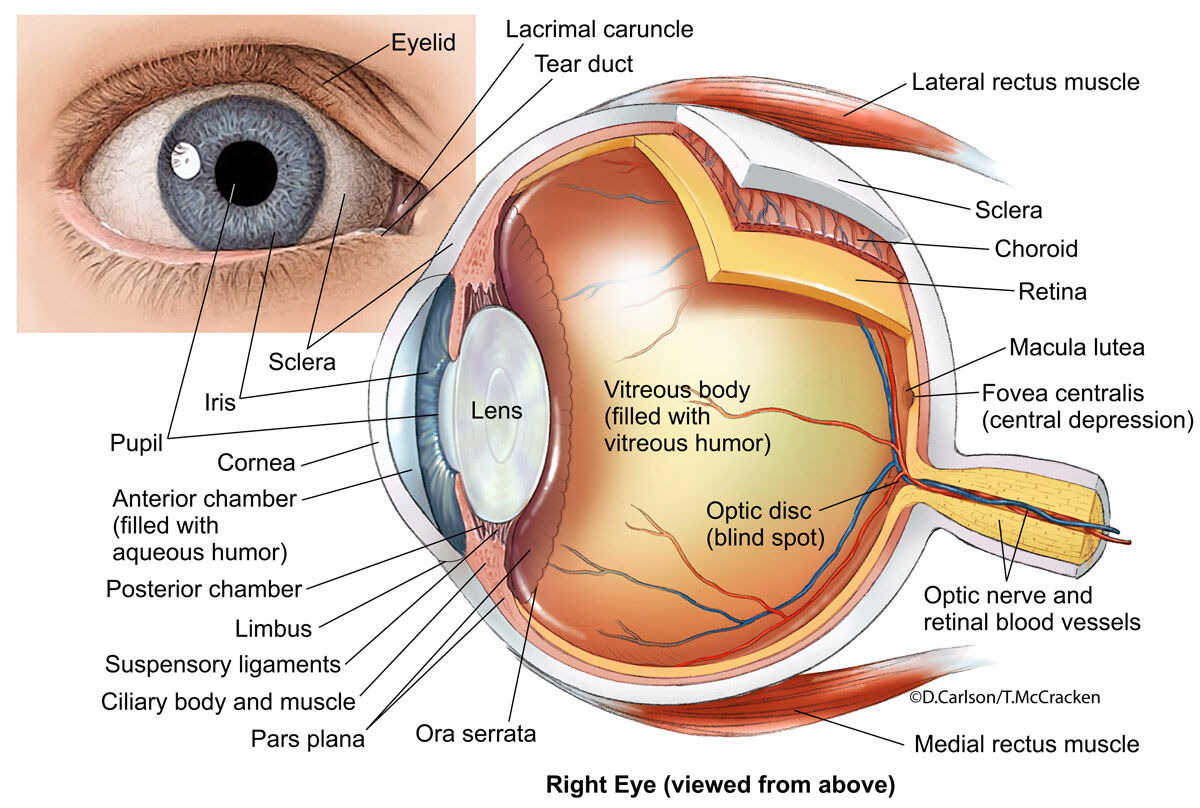
Extraocular muscles help move the eye in different directions.

External anatomy of the eye. The episclera is covered by the conjunctiva a vascular mucous membrane. Human eye parts 1. Anatomy of the eye.
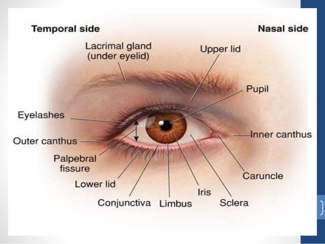
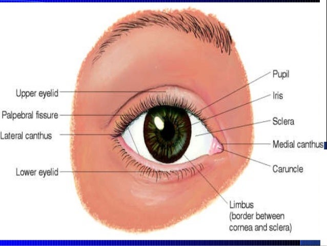
It is the most visible part of the eye. Squinting which is closing the eye partially sheilds the eye from excessive light that can damage the internal structure such as the retina. The outer margin of the cornea.
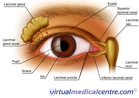
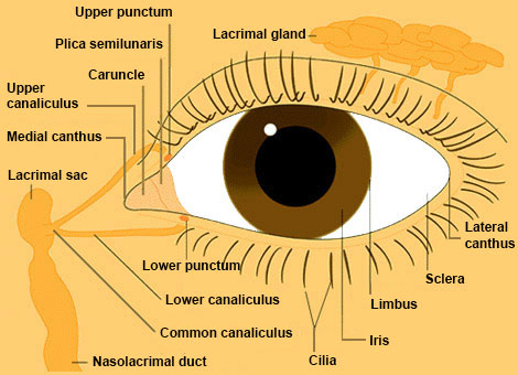
The human eye ball is spherical in structure and is about 24 mm in a diameter. Component of the lacrimal apparatus located superior and lateral to each eye. Overview the cornea allows light to enter the eye.
The outer fibrous or sclera 2. The diaphragm that controls the amount of light entering the eye. Component of the lacrimal apparatus tears enter thru lacrimal punctum.
As light passes through the eye the iris changes shape by expanding and letting more light through or constricting and letting less light through to change pupil size. Nerve signals that contain visual information are transmitted through the optic nerve to the brain. The eye is surrounded by the orbital bones and is cushioned by pads of fat within the orbital socket.
It has two layers a posterior pigment epithelium and an anterior stroma made of collagen muscle and pigment cells. Lacrimal system tear drainage system the lacrimal system is crucial for tear production and management which includes distribution of tears and draining excess tears. Malfunction in any part of the system can cause serious complications.
The lens then changes shape to allow the accurate focusing of light on the retina. The iris is the colored part of the eye that controls the amount of light that enters into the eye. It is the most visible part of the eye.
It lies in front of the crystalline lens and separates the anterior chamber from the posterior chamber.
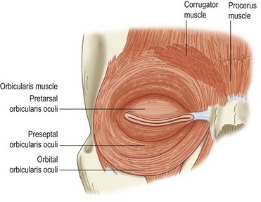 Eyelid Anatomy And Function Clinical Gate
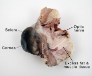 Cow Eye Dissection Anatomy Project Hst Learning Center
 Anatomy Of Eye
 Special Senses Vision Overview Of Special Senses
 Diagram Of The Eye Lions Eye Institute
 An Exploration Of The Eye Light Is Essential For Vision
External Anatomy Of The Eye Diagram Quizlet
 External Anatomy Of The Eye And Accessory Structures
 Vision And The Eye S Anatomy Healthengine Blog
 Ppt External Anatomy Of The Eye Powerpoint Presentation
A P Lab Exam 2 Study Guide Bmch 2400 Unomaha Studocu
 3 Eye External Anatomy Recorded Lecture Video Youtube
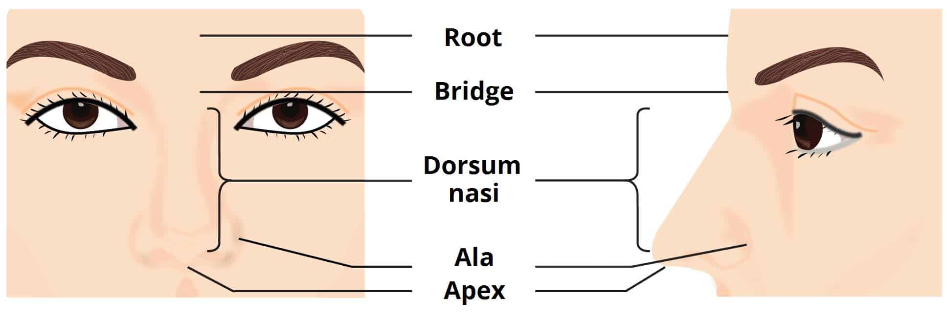 The External Nose Muscles Innervation Teachmeanatomy
 The Eye And Vision
 Special Senses Anatomy And Physiology Nurseslabs
 Eye Anatomy 2 Illustration Carlson Stock Art

 Goldfish External Anatomy Solid Gold Aquatics
 External Anatomy Of The Eye And Accessory Structures Diagram

 Eye Wikipedia
 Fish Anatomy Internal External Anatomy Of A Fish
 Eyes
Eyes
 671 02102584

 Human Eye Wikipedia
Stock Image Illustration Of The Normal External Anatomy Of

Belum ada Komentar untuk "External Anatomy Of The Eye"
Posting Komentar