Pulmonary Veins Anatomy
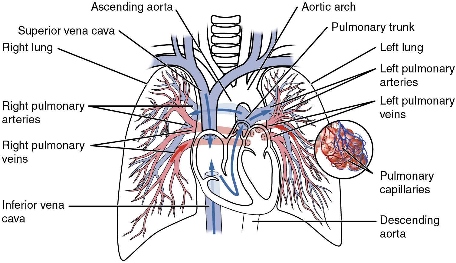
The pulmonary capillaries surround and embrace millions of tiny air sacs called alveoli in your lungs. The pulmonary veins pvs are large blood vessels that carry oxygenated blood from the lungs and drain into the left atrium la of the heart.
 20 5 Circulatory Pathways Anatomy And Physiology
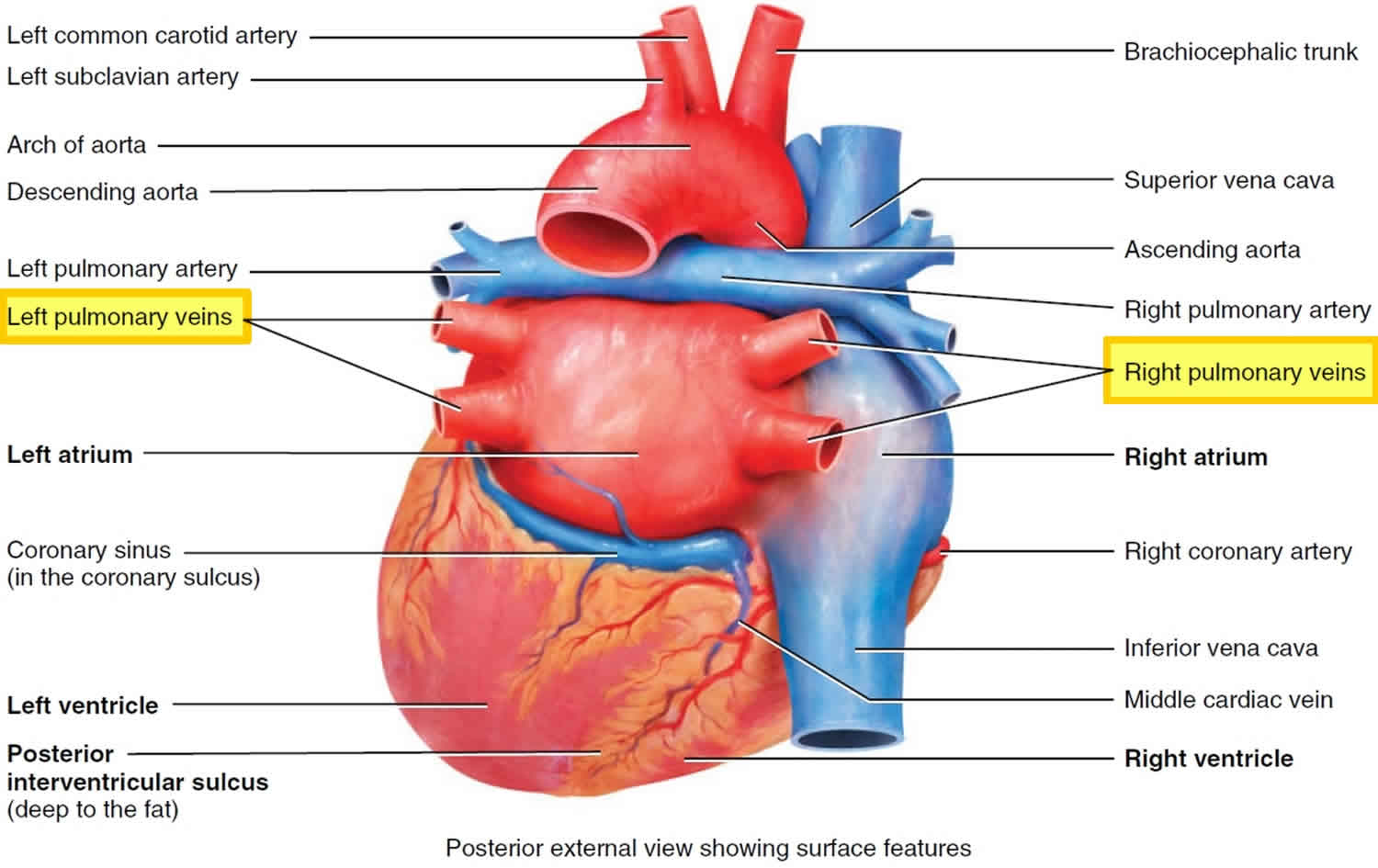
Most individuals have four pulmonary veins two on the left and two on the right the inferior and the superior one but there are also different anatomical variations.

Pulmonary veins anatomy. Typically there are four pulmonary veins with superior and inferior pulmonary veins on either side draining into the left atrium. The pulmonary veins along with the pulmonary arteries make up the pulmonary circulation. Drains the left upper lobe.
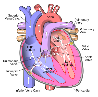
Just like the other veins in your body your pulmonary veins arise from a network of capillaries. Veins are the blood vessels that carry blood to the heart. Pulmonary veins are responsible for carrying oxygenated blood from the lungs back to the left atrium of the heart.
The pulmonary veins serve a very important purpose of delivering freshly oxygenated blood. Embryologically the pulmonary arteries originate from the truncus arteriosus. There are typically four pulmonary veins two draining each lung.
This differentiates the pulmonary veins from other veins in the body which are used to carry deoxygenated blood from the rest of the body back to the heart. Large veins have diameters greater. Drains the left lower lobe.
Anatomy of the pulmonary veins. While veins usually carry deoxygenated blood from tissues back to the heart in this case. However the capillaries that give rise to the pulmonary veins differ from capillaries elsewhere in your body.
The pulmonary veins are part of the pulmonary circulation. Histology of the arteries and veins. The pulmonary veins can be affected by.
Drains the right upper and middle lobes. The pulmonary veins are the veins that transfer oxygenated blood from the lungs to the heart. The distal segments of the pulmonary veins are intrapericardial.
Drains the right lower lobe. The largest pulmonary veins are the four main pulmonary veins two from each lung that drain into the left atrium of the heart. The anatomy of the pulmonary vein anatomy.
Pulmonary arteries and veins anatomy. The portion of myocardium extending onto the pulmonary veins is a frequent source of atrial fibrillation and the left superior pulmonary vein which has the longest segment of myocardium is the focal cause of atrial fibrillation in as many as half of the cases 12.
 What Do The Four Pulmonary Veins Empty Into Socratic

 Pulmonary Vein An Overview Sciencedirect Topics
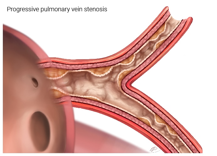
 Pulmonary Vein Stenosis
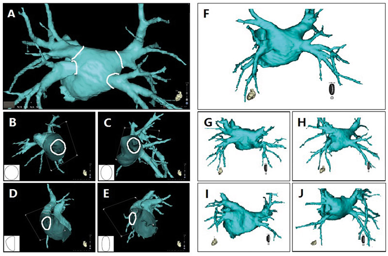
 Clinical Relevance Of Computed Tomography Pulmonary
 Anatomical And Functional Evaluation Of Pulmonary Veins In
 Pulmonary Vein Anatomy Function Location Ablation
Right Top Pulmonary Vein Draining The Posterior Segment Of
 A Usual Superoinferior Pulmonary Venous Arrangement At
 Cardiac Anatomy For The Electrophysiologist With Emphasis On
 Pulmonary Vein Wikipedia
 Main Bronchi With Pulmonary Arteries And Veins In Situ

 A Normal Pulmonary Venous Anatomy Volume Rendered Image Of
 Chest Radiology
 Anatomical And Electrophysiological Basis For Pulmonary Vein
 Role Of Pulmonary Veins In Initiation Of Atrial Fibrillation
File Left And Right Pulmonary Veins Anatomy Function
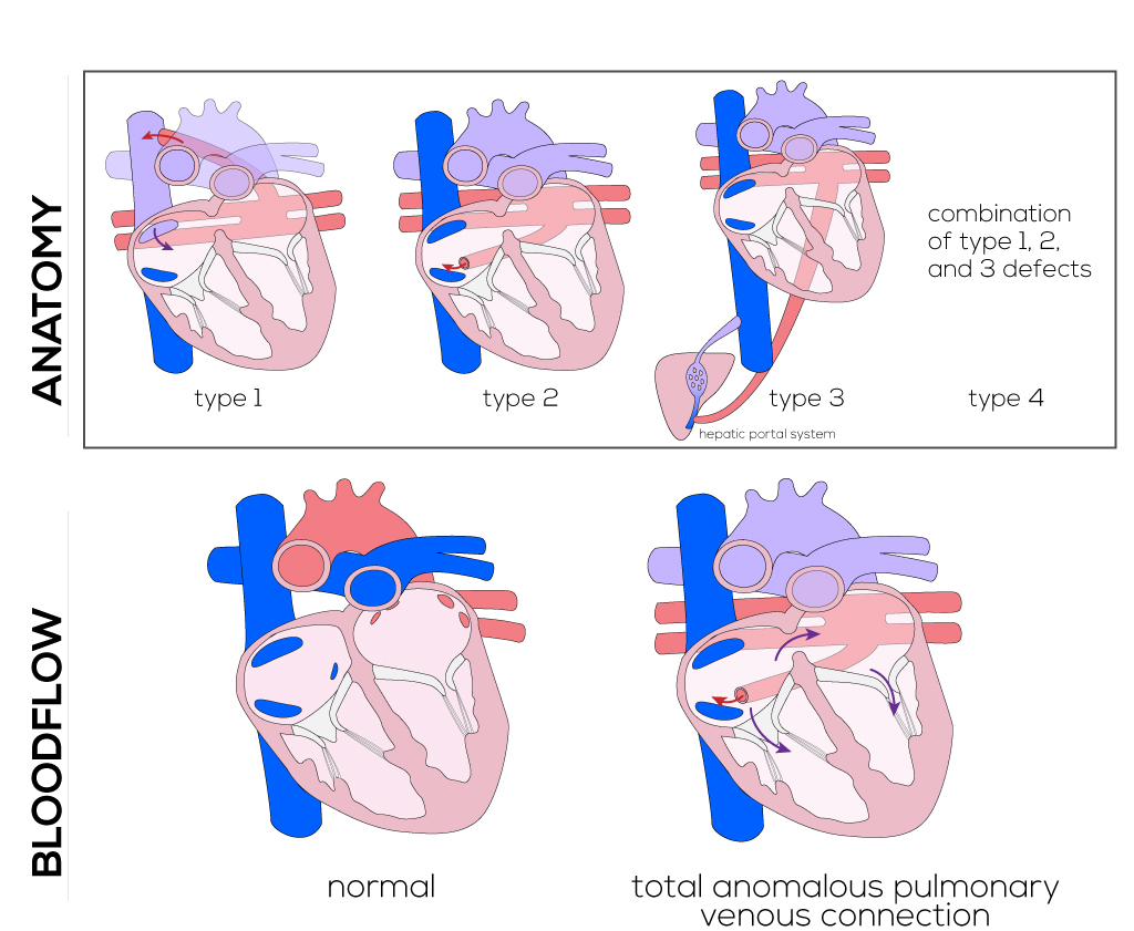
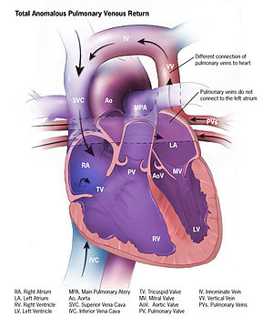 Anomalous Pulmonary Venous Connection Wikipedia
 Anatomy Of The Respiratory System 6
 Anatomical Characteristics Of The Junction Of Left Atrium
 Anatomical Regions Of The Left Atrium La And Pulmonary
 Electrophysiology Of Pulmonary Veins Venous And Lymphatic
Belum ada Komentar untuk "Pulmonary Veins Anatomy"
Posting Komentar