The Tooth Anatomy
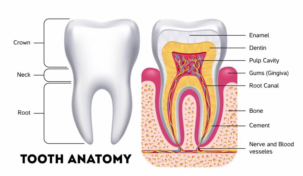
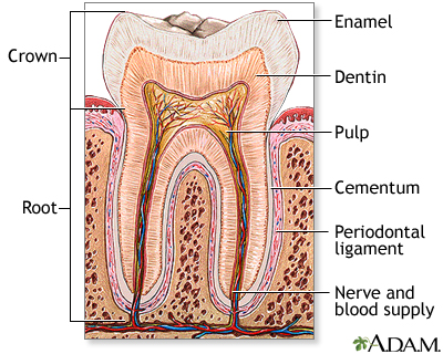
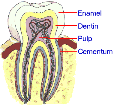
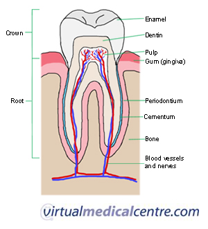
It is a hard tissue that contains microscopic tubes. The enamel dentin cementum and pulp.
 The Anatomy Of A Tooth In Four Parts Arc Dental
Tooth anatomy types of teeth.
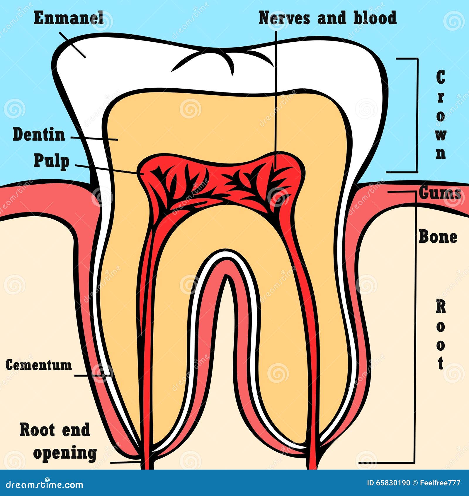
The tooth anatomy. Enamel this is the outer and hardest part of the tooth that has the most mineralized tissue in. Each tooth has several distinct parts. The hardest white outer part of the tooth.
Anatomy of the tooth the tooth is one of the most individual and complex anatomical as well as histological structures in the body. The tooth is made of several layers of varying density and hardness. The tissue composition of a tooth is only found within the oral cavity and is limited to the dental structures.
A layer of connective tissue that binds the roots of the. Dental anatomy is a field of anatomy dedicated to the study of human tooth structures. The enamel is the outer layer of teeth that covers the crown.
The softer living inner structure of teeth. A layer underlying the enamel. Teeth are used for catching and masticating food for defense and for other specialized purposes.
The root is the part of the tooth that extends into the bone and holds. Dental anatomy is also a taxonomical science. Tooth anatomy the structure of a human tooth includes the following tissues.
Enamel the hard calcified outer covering which is used to break down food. Most people start off adulthood with 32 teeth not including the wisdom teeth. Tooth formation begins before birth and the teeths eventual morphology is dictated during this time.
Picture of the teeth enamel. Tooth enamel is the most resistant and hardest tissue in the human body. Tooth plural teeth any of the hard resistant structures occurring on the jaws and in or around the mouth and pharynx areas of vertebrates.
Pulp this is the soft tissue found in the center of all. The soft tissue found at the centre of the tooth in both crown and root pulp contains nerves and blood vessels which alow the tooth to feel pain and sensation. Here is an overview of each part.
Usually there are 20 primary te. Enamel consists primarily of a matrix of hydroxyapatite a mineral made of crystalline calcium phosphate which is created by the bodys cells during tooth development. Also it protects the tooth against external damaging forces.
It is concerned with the naming of teeth and the structures of which they are made this information serving a practical purpose in dental treatment. Dentin this is the layer of the tooth under the enamel. Explore the interactive 3 d diagram below to learn more about teeth.
The development appearance and classification of teeth fall within its purview.
Dental Cavities In Children Lonestar Smiles For Kids
 Periodontium Or The Tooth Supporting Structure
 Free Anatomy Quiz The Teeth Quiz 1
 Tooth Anatomy Structure Function With Pictures
 Tooth Anatomy Medlineplus Medical Encyclopedia Image
 Tooth Anatomy Tissues Of A Tooth
 Tooth Anatomy Scheme Stock Illustration Illustration Of
:background_color(FFFFFF):format(jpeg)/images/library/12317/the-anatomy-of-the-tooth_english__1_.jpg) Tooth Anatomy Structure Parts Types And Functions Kenhub
Tooth Anatomy Structure Parts Types And Functions Kenhub
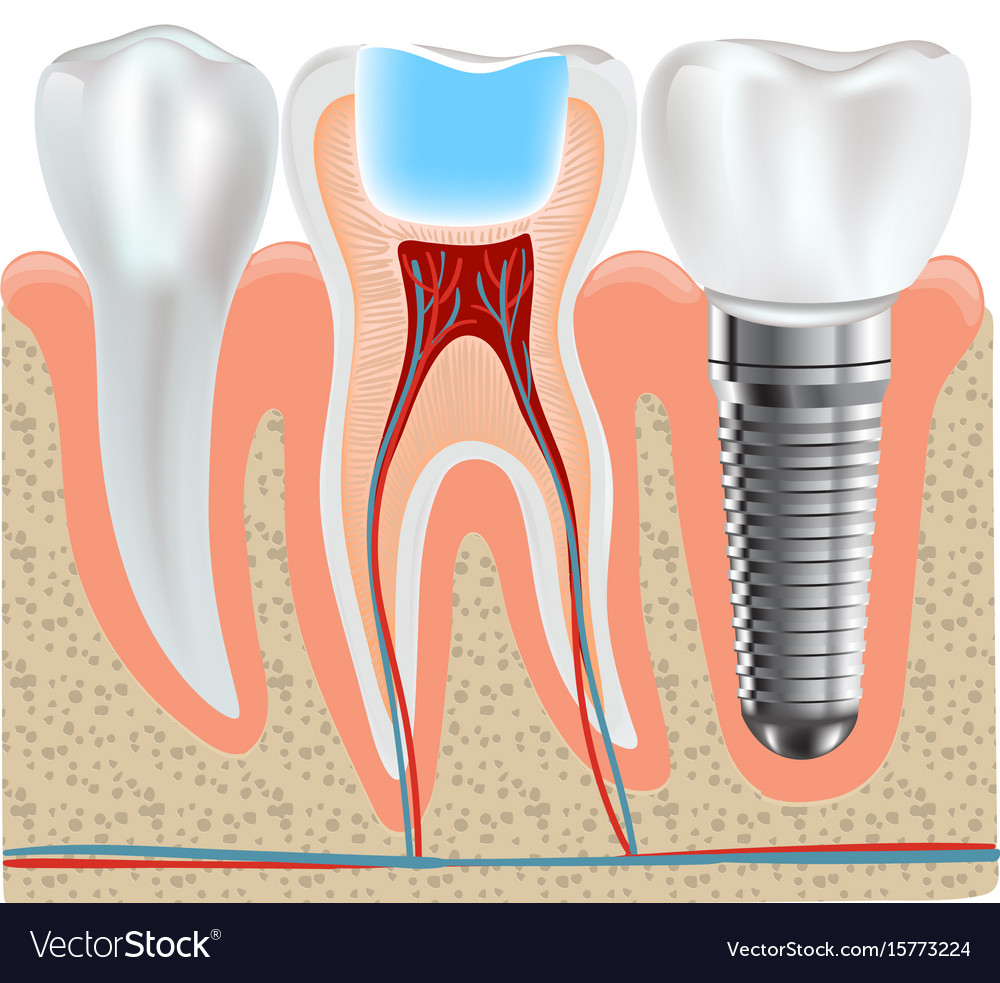 Dental Implant And Real Tooth Anatomy Closeup
 Information About The Human Tooth Anatomy With Labeled
Vc Dental Tooth Anatomy Education
 Tooth American Dental Association
 Tooth Anatomy Infographics Realistic White Tooth
 1 General Tooth Anatomy Indicating The Four Primary Types
 Tooth Anatomy Everything You Should Know About The Teeth
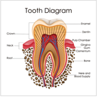 A Look At The Anatomy Of A Tooth Wake Orthodontics
 Oral Care Dental Assistant Study Dental Hygiene Student
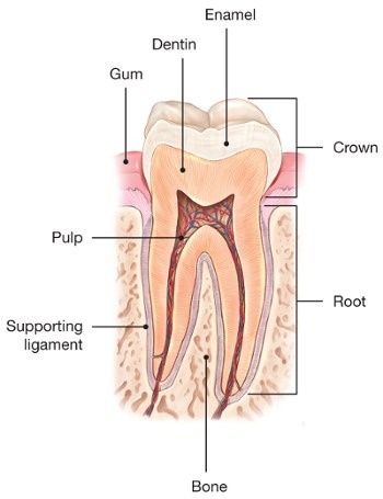 Cracked Teeth American Association Of Endodontists
Painful Tooth Treatment Anatomy Of The Tooth Art
The Anatomy Of A Tooth Oral Answers
 Anatomy Of The Teeth Malmin Dental Blog
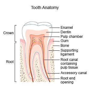 Hypomineralization Of The Tooth What You Need To Know
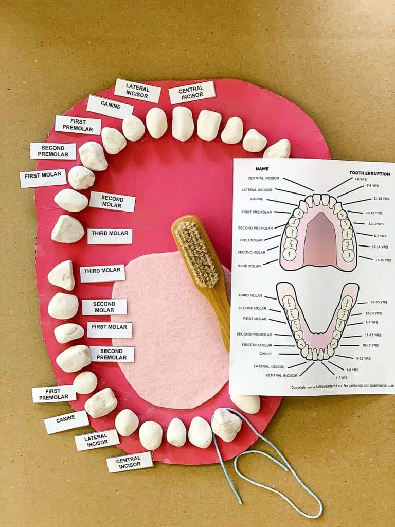 Teeth Mouth Anatomy Learning Activity Hello Wonderful
 Teeth Anatomy Adult Teeth Permanent Dentition
 Divisions And Components Of The Teeth An Overview Of


/toothanatomy2.jpg)


Belum ada Komentar untuk "The Tooth Anatomy"
Posting Komentar