Third Ventricle Anatomy
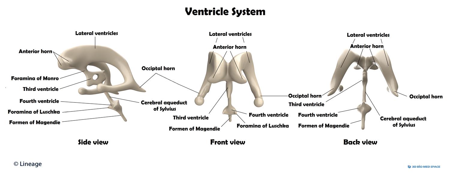
Third ventricle ventriculus tertius the third ventricle is a narrow funnel shaped unilocular midline cavity. The lateral ventricles are connected to the third ventricle by the foramen of monro.
 Surgery In And Around The Brain Stem And The Third Ventricle Anatomy Pathology Neurophysiology Diagnosis Treatment
The third ventricle is one of the four csf filled cavities that together comprise the ventricular system.

Third ventricle anatomy. The third ventricle is a narrow cavity that is located between the two halves of the brain. The third ventricle therefore serves as the intermediary between the lateral ventricles and the fourth ventricle. As with the other ventricles of the brain it is filled with cerebrospinal fluid which helps to protect the brain from injury and transport nutrients and waste.
It is connected at the superior anterior corner to the lateral ventricles by the foramina of monro and becomes the cerebral aqueduct. Slit like space lying in the sagittal plane it communicates at its anterosuperior margin with each lateral ventricle through the foramen of monro and posteriorly with the fourth ventricle through the aqueduct of sylvius. Anatomy of third ventricle it location structures forming boundaries recesses and tela choroidea and choroid plexus of third ventricle.
The third ventricle receives csf from the lateral ventricles and conveys it to the fourth ventricle which disseminates it to the subarachnoid space. The third ventricle is situated in between the right and the left thalamus. The third ventricle is one of the four csf filled cavities that together comprise the ventricular system.
The third ventricle is situated in between the right and the left thalamus. The third ventricle helps to protect the brain from trauma and injury. Of silvius at the posterior caudal corner.
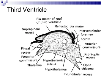
The third ventricle is a narrow laterally flattened vaguely rectangular region filled with cerebrospinal fluid and lined by ependyma. The third ventricle is a median cleft between the two thalami and is bounded laterally by them and the hypothalamus. The third ventricle is a median cleft between the two thalami and is bounded laterally by them anteriorly and the hypothalamus and subthalamus posteriorly.
It is a cavity filled with cerebrospinal fluid located between the two hemispheres of the diencephalon of the forebrain. Its anterior wall is formed by the lamina terminalis and posteriorly there is the pineal recess. The third ventricle is one of four brain ventricles.
The third ventricle is one of the four ventricles in the brain that communicate with one another.
 Schematic Drawing Of The Related Nuclei Of The Third
 Walls Of The Third Ventricle Anatomical Illustration Art Print
 Endoscopic Anatomy Of The Third Ventricle Microsurgical And
 Figure 12 From The Cerebrum Anatomy Semantic Scholar
:max_bytes(150000):strip_icc()/third_ventricle-57ed3f703df78c690f801ee9.jpg) Third Ventricle Function And Anatomy
Third Ventricle Function And Anatomy
 Endoscopic Third Ventriculostomy Pacific Adult
:watermark(/images/watermark_only.png,0,0,0):watermark(/images/logo_url.png,-10,-10,0):format(jpeg)/images/anatomy_term/plexus-choroideus-ventriculi-tertii/k3Kar8lR1xxFD9LkkAKwXA_image1_medial.png) Third Ventricle Anatomy Kenhub
Third Ventricle Anatomy Kenhub
 Ventricular Anatomy Postgraduate Training
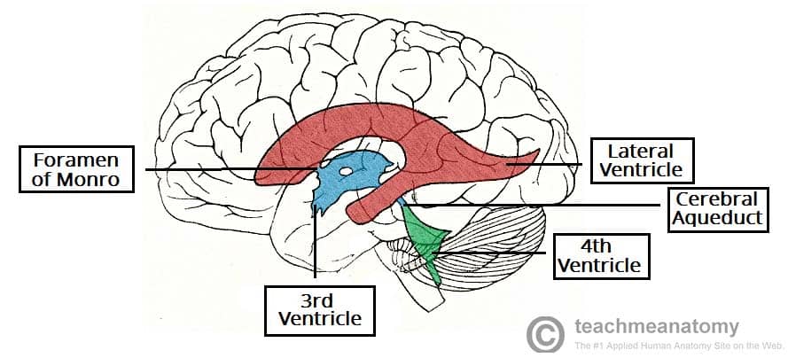 The Ventricles Of The Brain Lateral Third Fourth
 Third Ventricle An Overview Sciencedirect Topics
 Third Ventricle Surgical Anatomy And Approaches
Overview Of The Central Nervous System Gross Anatomy Of The
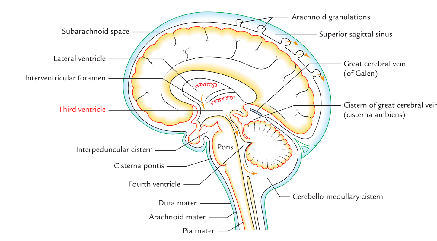 Easy Notes On Third Ventricle Learn In Just 4 Minutes
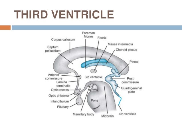 Third Ventricle Surgical Anatomy And Approaches
 Histological Analysis Of The Third Ventricle Floor In
 Lateral Ventricles An Overview Sciencedirect Topics
 Walls Of The Third Ventricle Anatomical Illustration Poster
 Endoscopic Third Ventriculostomy An Overview
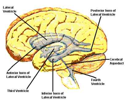 Third Ventricle Medfriendly Com
:max_bytes(150000):strip_icc()/brain_ventricles-56d0ccd03df78cfb37b876dc.jpg) Ventricular System Of The Brain
Ventricular System Of The Brain
 Ventricles And Coverings Of The Brain Clinical
 Cerebrospinal Fluid Csf Neurology Medbullets Step 1
 Third Ventricle Boundaries Ranzcrpart1 Wiki Fandom
 A P Final 2 Brain At North Shore Community College Studyblue
 Neuroanatomy Online Lab 3 The Ventricles And Blood Supply


Belum ada Komentar untuk "Third Ventricle Anatomy"
Posting Komentar