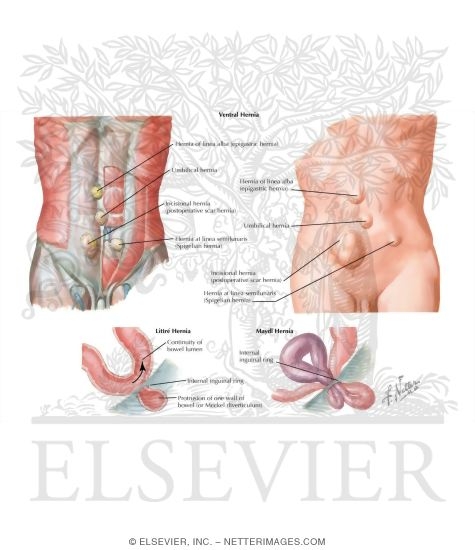
Ventral Hernia Anatomy
Many are called incisional hernias. Abdominal wall hernias and relevant surgical techniques.
 Abdominal Hernias Treatment Management Approach
Ventral hernia repair is surgery to repair a ventral hernia.

Ventral hernia anatomy. Uncommon in other animals abdominal wall hernias are among the most common. The cause of a ventral hernia can differ depending on its location as well as your medical history health and personal anatomy. The most common types of hernia are inguinal inner groin.
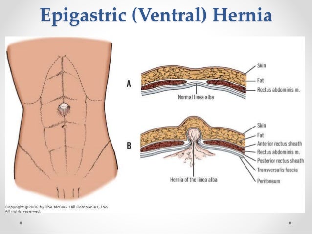
Ventral hernias often occur at the site of an old surgical cut incision. The majority of hernias form in the weaker areas of the abdominal wall where there is no muscle present and typically occur in the linea alba. A ventral hernia is a sac pouch formed from the inner lining of your belly abdomen that pushes through a hole in the abdominal wall.
Umbilical and incisional hernias are specific types of ventral hernias. As the adjective suggested it referred to a particular section being trapped or confined. These layers are a bit different between the umbilical region and the groin but overall the basic layers are the same.
A ventral hernia is a disruption or hole in the abdominal wall and can be classified as primary occurring de novo or incisional hernias caused by previous incision and surgery. The pinchcock action of the internal ring musculature during abdominal muscular. When you have an incarcerated ventral hernia it poses far more than some pains and a sore abdomen.
A ventral hernia is a bulge of tissues through an opening of weakness within your abdominal wall muscles. This type of ventral hernia is also called incisional hernia. A hernia occurs when there is a hole in the muscles of the abdominal wall allowing a loop of intestine or abdominal tissue to push through the muscle layer.
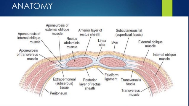
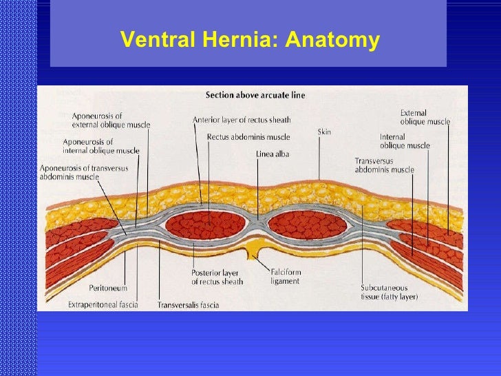
A hernia occurs when an organ or fatty tissue squeezes through a weak spot in a surrounding muscle or connective tissue called fascia. It can occur at any location on your abdominal wall. Hernia anatomy the layers of the abdominal wall the first concept to understand is the basic layers of the abdominal wall.
The anterior abdominal wall is composed of multilaminar mirror image muscles. A ventral hernia is a hernia that occurs at any location along the midline vertical center of the abdomen wall. There are different locations and kinds of hernia which requires individual management.
What is a ventral hernia. A ventral abdominal hernia refers to any protrusion of intestine or other tissue through a weakness or gap in the abdominal wall. However certain ventral hernias may be seen in any location at the abdomen.
Ventral hernias are one of the classifications of abdominal hernia that occurs along the anterior midline portion of the abdomen. When a weakened pocket of the abdominal wall traps your intestine where it cant be pushed back inside your cavity blockage may occur.
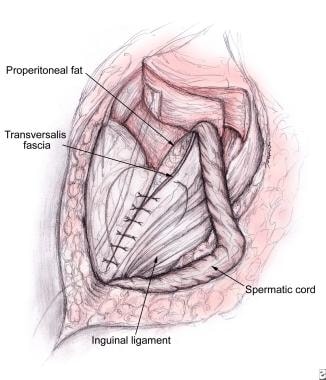
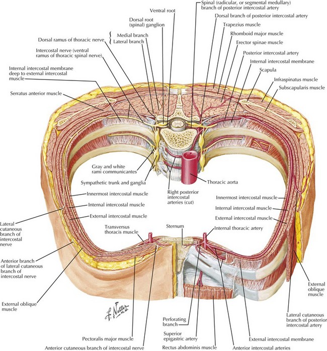
 Surgical Anatomy Of Anterior Abdominal Wall
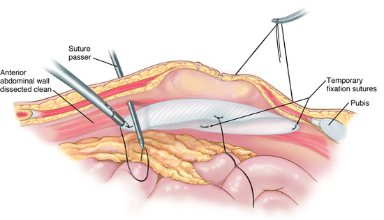
 Laparoscopic Ventral Hernia Repair Information From Sages
 Hernias Tintinalli S Emergency Medicine A Comprehensive
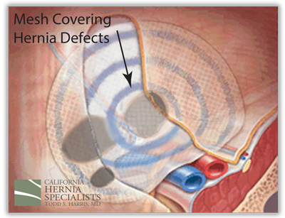 Hernia Anatomy California Hernia Specialists

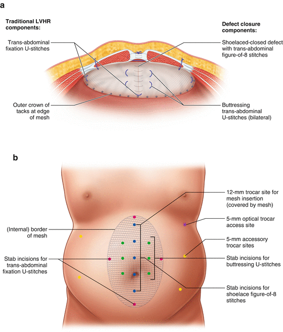 Laparoscopic Ventral Hernia Repair With Defect Closure

 Lap Ventral Hernia Surgery In Indore Vijay Nagar By
 Ventral Hernia Repair Medical Illustration Human Anatomy
 Division Of The Posterior Rectus Sheath A And
 Abdominal Wall Ventral Hernias
 Abdominal Hernia Anatomy Of Female Lateral View Medical
 Abdominal Wall Hernias
Stock Image Illustration Of An Incisional Ventral Abdominal
 Ventral Hernia Mesh Repair Gallbladder Surgery Umbilical
 Laparoscopic Ventral Hernia Repair Medical Exhibit Medivisuals
 Doctors For Hernia Surgery In Chennai Credihealth
Ventral Hernia Artwork Stock Image C020 6693 Science
 Ventral Hernia Repair Medical Illustration Human Anatomy
 Several Techniques Of Mesh Repair For Incisional Or Ventral
 Component Seperation Technique For The Repair Of Very Large
 Ventral Hernia Repair Medical Illustration Human Anatomy
 Ventral Hernia General Surgery Surgical Anatomy Original Illustration Pen And Ink 6x9 In
 The Endoscopic Retromuscular Repair Of Ventral Hernia The
 Ventral Hernia Challenges And Choices
 Science Source Ventral Hernia
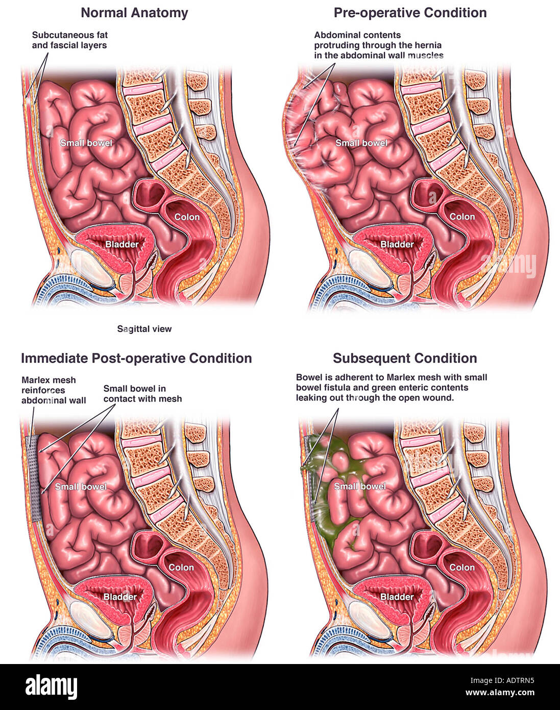 Ventral Hernia Repair With Post Operative Complications
 Open Ventral Hernia Repair Basicmedical Key
Belum ada Komentar untuk "Ventral Hernia Anatomy"
Posting Komentar