Anatomy Of The Knee Cap
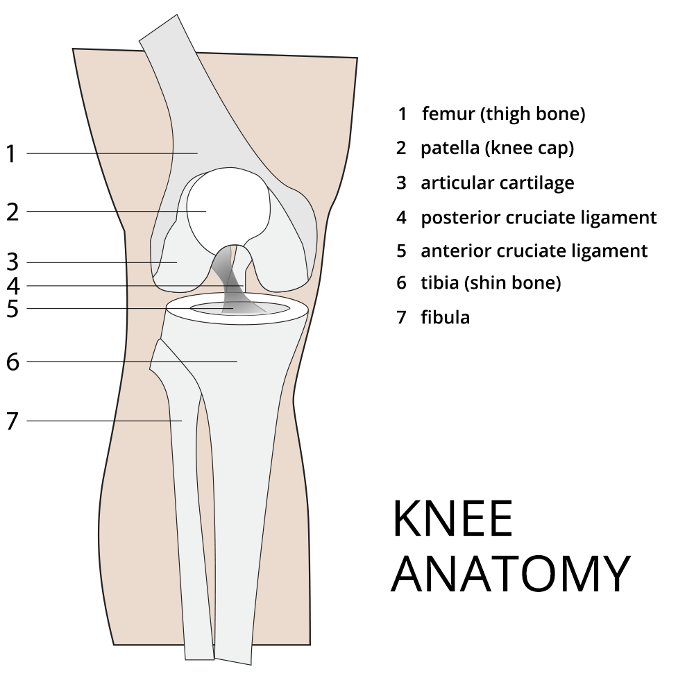
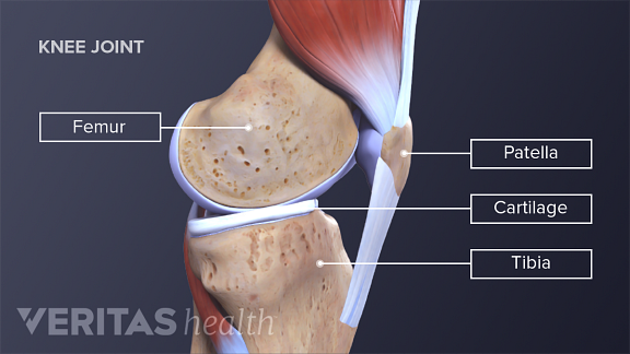
It protects the bones and soft tissue in your knee joint and slides when your knee moves allowing leverage in your leg muscles. The femur thigh bone tibia shin bone patella knee cap fibula smaller bone next to shin bone.

Muscles tendons and ligaments connect the knee bones.
Anatomy of the knee cap. Acts as a shock absorber. Femur the upper leg bone or thigh bone. Makes walking more efficient.
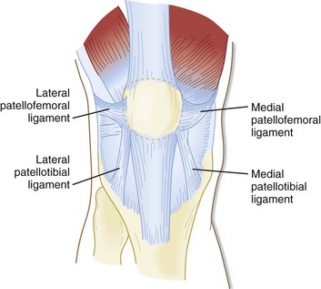
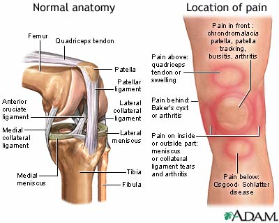
The kneecap glides in a groove in the thighbone and adds leverage to the thigh muscles which are used to extend the leg. The smaller bone that runs alongside the tibia fibula and the. Two concave pads of cartilage strong flexible tissue called menisci minimize the friction created at the meeting of the ends of the tibia and femur.
There are also several key ligaments a type of fibrous connective tissue that connect these bones. The knee is designed to fulfill a number of functions. Helps to lower and raise the body.
The largest joint in the body the knee moves like a hinge allowing you to sit squat walk or jump. The knee joins the thigh bone femur to the shin bone tibia. Bones of the knee.
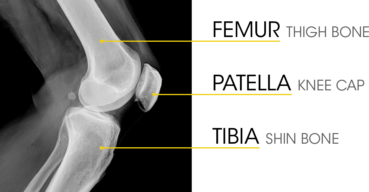
Allows twisting of the leg. The knee is one of the largest and most complex joints in the body. The knee consists of three bones.
Tibia the bone at the front of the lower leg or shin bone. Support the body in an upright position without the need for muscles to work. The knee is the joint where the bones of the lower and upper legs meet.
Patella knee cap the patella is the larger sesamoid bone in your body and rests over a groove at the bottom of the femur and the top of the tibia. Another bone the patella kneecap is at the center of the knee. Helps propel the body forward.
Adolescent Sports Injuries Of The Knee Cleveland Clinic
 Patellofemoral Stress Syndrome Towson Orthopaedic Associates
 Knee Joint Anatomy Bones Cartilages Muscles Ligaments
Soft Tissue Knee Patient Information Gavin Mchugh
 Anatomy Of Human Knee Joint Art Print
 Patellofemoral Joint Physiopedia
 Matthew Boyle Orthopaedic Surgeon Knee Anatomy Knee
 Prepatellar Bursitis Orthopedic Knee Specialist Richmond Va
 The Knee Anatomy Injuries Treatment And Rehabilitation
 Knee Pain Medlineplus Medical Encyclopedia
 Muscles Of The Knee Anatomy Pictures And Information
:max_bytes(150000):strip_icc()/vector-illustration-of-a-meniscus-tear-and-surgery-871162428-03ac23d73f854954a8082f2ae3ce9219.jpg) Meniscus Vs Cartilage Tear Of The Knee
Meniscus Vs Cartilage Tear Of The Knee
:background_color(FFFFFF):format(jpeg)/images/library/5745/ssCW1ZC0OK3HY0uiT407Q_Patella_01.png.jpeg) Patella Anatomy Function And Clinical Aspects Kenhub
Patella Anatomy Function And Clinical Aspects Kenhub
 Anatomy Of The Knee Central Coast Orthopedic Medical Group
 Understanding The Role Of Cartilage In The Knee
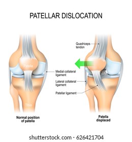 Patella Dislocation Images Stock Photos Vectors
Common Knee Injuries Orthoinfo Aaos
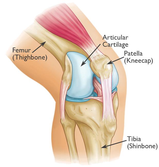 Patellar Fractures Broken Kneecap Orthoinfo Aaos
 Anatomy Of Patella Bone And Spine
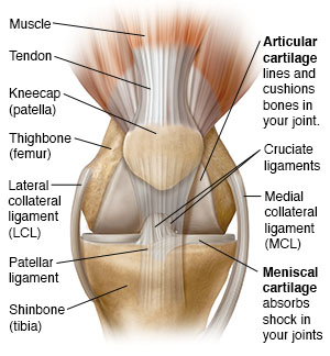
 Runner S Knee Part 1 What Is Patellofemoral Pain Syndrome
 Knee Human Anatomy Function Parts Conditions Treatments
Functional Anatomy Of The Knee Movement And Stability
 Anatomy Of The Knee For Dancers Dance Work Balance
 Anatomy Of The Knee Baxter Regional Medical Center
 Knee Joint Picture Image On Medicinenet Com
 Luxating Patella Dislocated Knee Cap The Veterinary
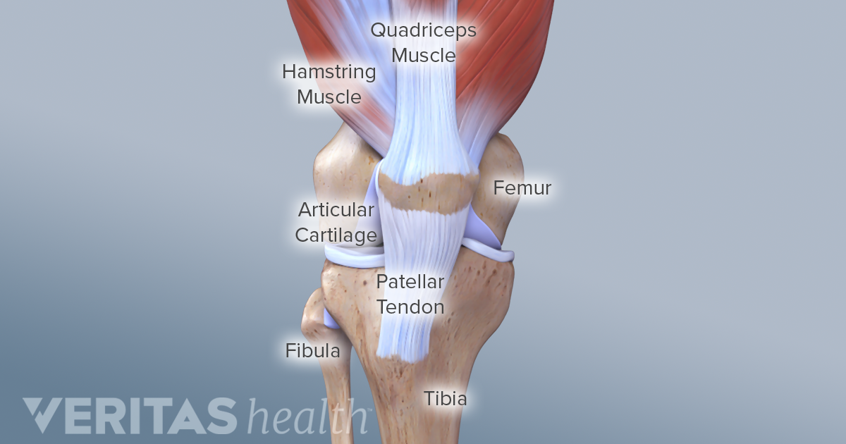



Belum ada Komentar untuk "Anatomy Of The Knee Cap"
Posting Komentar