Anatomy Of Fetal Heart
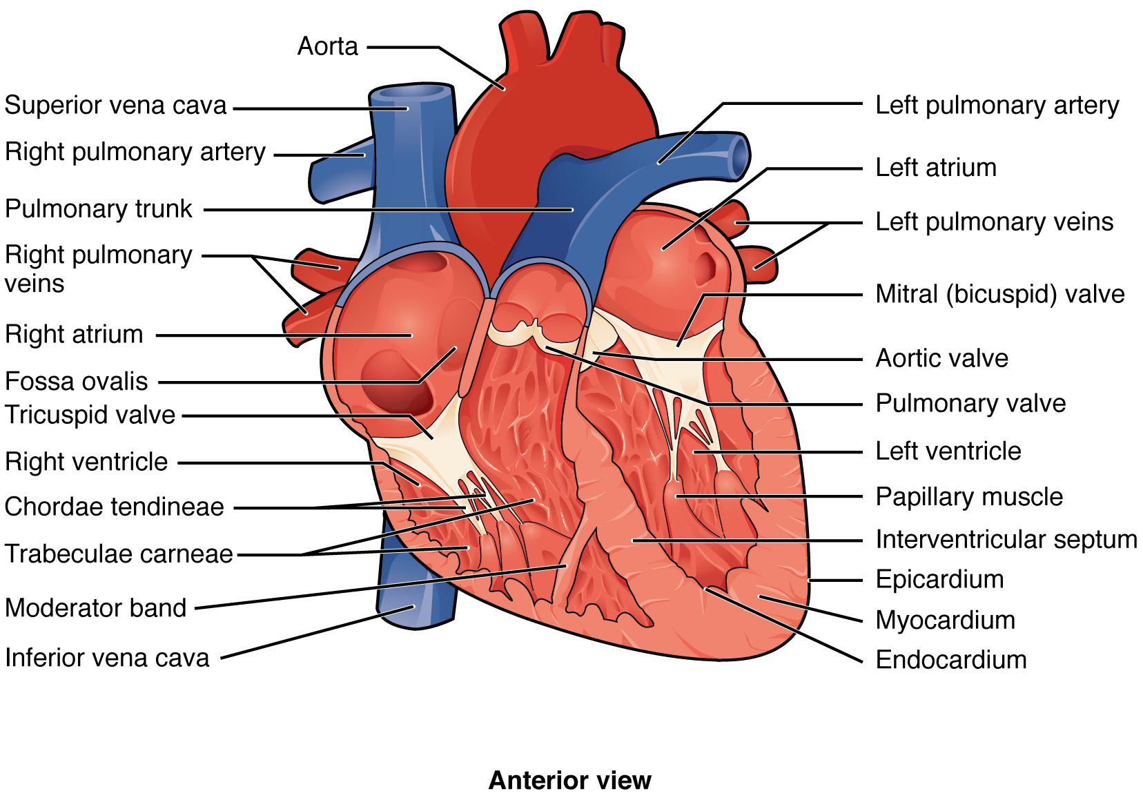
The channel closes postnatally becoming the arterial ligament. 5012 there are four chambers of the heart two atria in communication via a patent foramen ovale and two ventricles with complete septationthe left ventricular myocardium is configured differently from the right such that it is capable of working under higher pressure afterload.
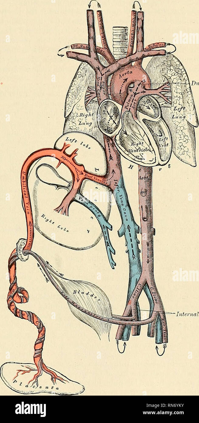
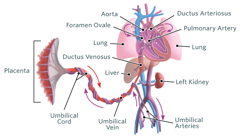
 Blood Circulation In The Fetus And Newborn Children S
The fetal heart and circulation at the end of 9 weeks are completely formed phoon 2001as depicted in fig.

Anatomy of fetal heart. Once the main arteries and veins as well as the heart are developed usually after the 8th week of fetal development deoxygenated blood is returned from the fetal systemic circulation to the placenta via two umbilical arteries which branch off the fetal internal iliac arteries. Home academics biology resources biology lab 107 resources histology anatomy of fetal pig thoracic cavity histology anatomy of fetal pig heart heart the heart is located in the thoracic cavity nestled between the lungs on the bodys midline. Mere presence of the heart in the location as described above and the finding of normal arrangement of the remaining organs however does not guarantee cardiac normality.
During fetal life in the absence of gas exchange the lungs receive only a small amount of blood volume pumped from the rv. The right side of the heart consists of the superior and inferior vena cava the right atrium the right ventricle. Ultrasound image of the right ventricular outflow tract rvot ultrasound images of the lvot left ventricular outflow tract some more images of lvot and rvot.
Fetal blood vessels and fetal circulation. Start a free trial of quizlet plus by thanksgiving lock in 50 off all year try it free ends in 02d 05h 28m 23s. In terms of its size the normal heart occupies about 13 of the fetal thorax and its transverse diameter of the heart must not exceed half of the transverse diameter of the chest.
Confirm that the heart is on the fetal left. Patency of the arterial duct or ductus arteriosus derived from the left sixth pharyngeal arch artery is a characteristic feature of the fetal heart. Normal sonographic anatomy of the fetal heart.
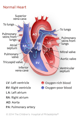
The left side of the heart consists of the pulmonary veins left atrium left ventricle and aorta. The heart should be angled 45degrees to the left and occupy approximately 13 of the chest. Fetal circulation after birth the circulation is divided into two separate sides that are not connected.
Choose from 500 different sets of fetal heart anatomy flashcards on quizlet. Contents of this page. Normal 3 vessel view in a 28 week fetus.
 Anatomy Descriptive And Applied Anatomy The Heart 569 The
 Fetal Heart Diagram Reading Industrial Wiring Diagrams
 19 1 Heart Anatomy Anatomy And Physiology
 Anatomy Of The Normal Fetal Heart The Basis For
 How Fetal Heart Work Fetal Circulation Medical Study
 Heart Anatomy Anatomy And Physiology
 Blood Circulation In The Fetus And Newborn Children S
 Fetal Heart Rate Tracing Physiology Fetal Heart Rate
 Pacific Medical Training Acls Bls Pals Certification
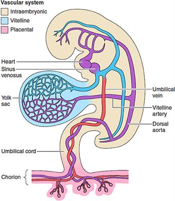 Frontiers Development Of The Human Placenta And Fetal
 Key Screening Views Of The Fetal Heart Part 3 4 Chamber View
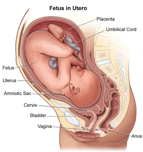 External And Internal Heart Rate Monitoring Of The Fetus
 Openstax Anatomy And Physiology Ch19 The Cardiovascular
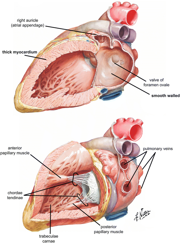 Anatomy Of The Human Heart Springerlink
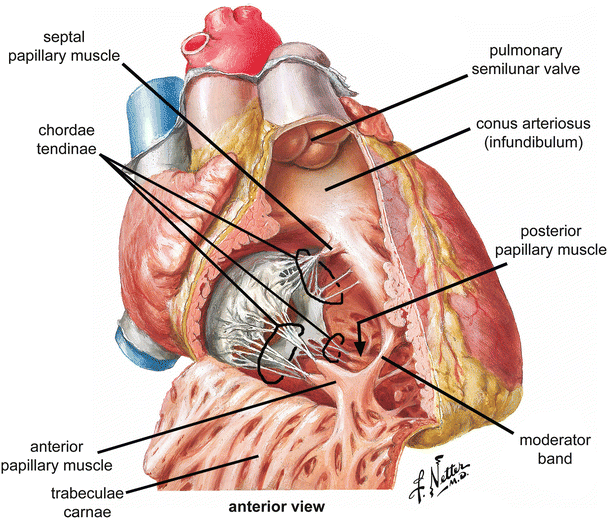 Anatomy Of The Human Heart Springerlink
 The Anatomy Ultrasound Everything You Should Know
Fetal Heart Anatomical Products
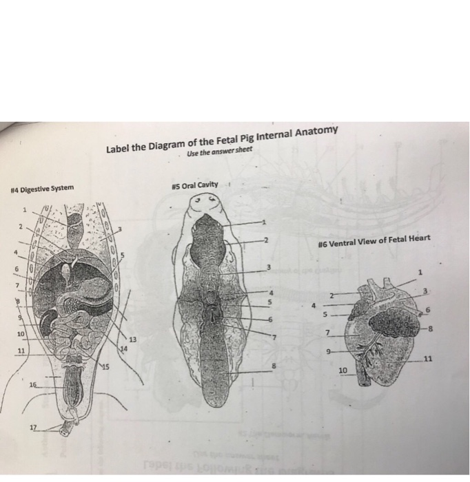 Solved Label The Diagram Of The Fetal Pig Internal Anatom
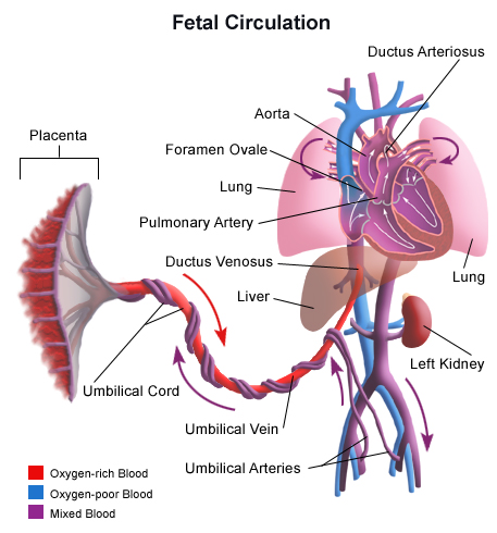
 6 Fetal Heart Anatomy And Electrical Activation Sequence
 Us 212 0 Anatomical Fetus Model With Viscus Placenta And Umbilical Cord Vivid Fetal Model With Heart Lung In Medical Science From Office School

Belum ada Komentar untuk "Anatomy Of Fetal Heart"
Posting Komentar