Peroneal Tendon Anatomy
Peroneal tendonitis can either be acute meaning that it comes on. There are three primary disorders of the tendons.
The muscles are connected to bone by the tendons.

Peroneal tendon anatomy. Peroneal tendon tears and tendonitis. Whats to know about peroneal tendonitis. The peroneal muscles are a group of two muscles of the leg.
People who take part in a sport that involves repetitive ankle motion are most prone. The occurrence of injuries to the peroneal tendons is not actually known. Anatomy origins insertions.
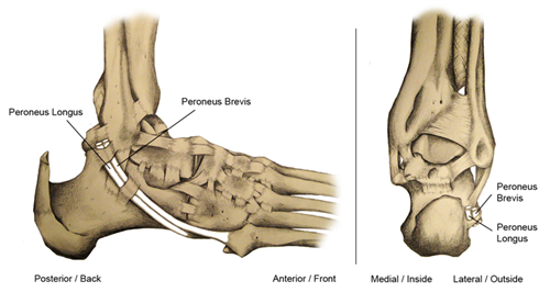
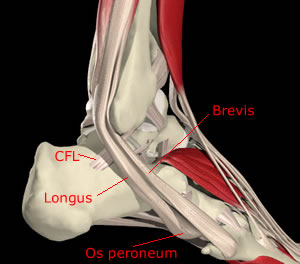
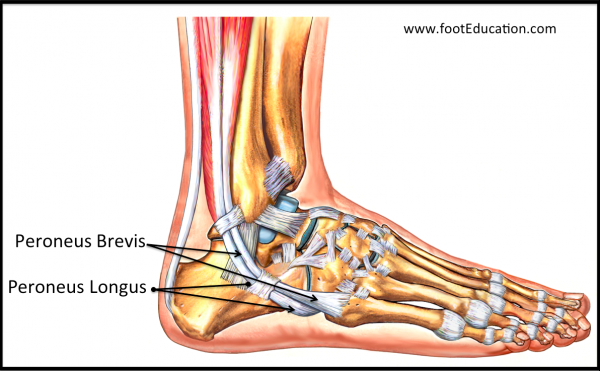
The peroneal tendons are located in the foot attaching muscle to bone. Normal variant anatomy in this region may include a peroneus quartus muscle a low lying peroneus brevis muscle belly or an os peroneum. At the level of the peroneal tubercle of the calcaneus.
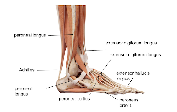
Peroneal tendon syndromes practice essentials. The two major peroneal muscles peroneus longus and peroneus brevis are situated on the outside of the leg just adjacent to the calf muscles. Fluid is seen in the common peroneal tendon sheath black.
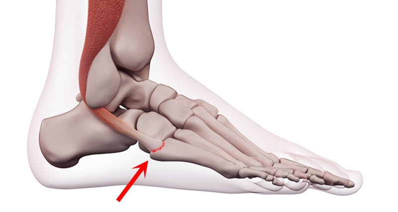
The peroneal tendons curved arrow are in their normal position within a convex fibular groove but thinning and irregularity of the peroneal brevis tendon suggest impending longitudinal splits. When lowering the foot they can be easily seen forming the surface of the lateral leg. The muscle the longest and most superficial of the three peroneus muscles is attached proximally to the head of the fibula and its belly runs down most of this bone.
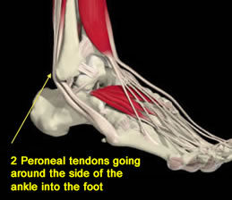
The peroneal tendons originate in the lateral compartment of the leg. In human anatomy the peroneus longus also known as fibularis longus is a superficial muscle in the lateral compartment of the leg and acts to evert and plantarflex the ankle. The peroneal tendons are the tendons that connect the muscles of the outer side of the calf to the foot.
Peroneal tendon disorders are a cause of hindfoot and lateral foot pain. These conditions are a cause of lateral ankle pain and may lead to ankle instability. The peroneal tubercle is variable in size and projects laterally from the anterior process of the calcaneus separating the positions of the peroneus brevis and longus tendons figure f.
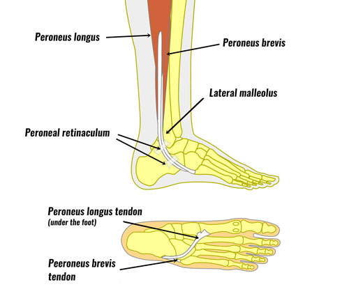
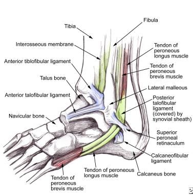
The sheath is runs in the retromalleolar sulcus on the fibula. Peroneal muscles of the leg. They lie within the peroneal compartment located at the lateral fibular region.
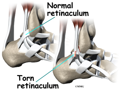
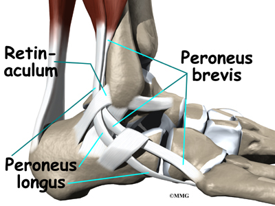
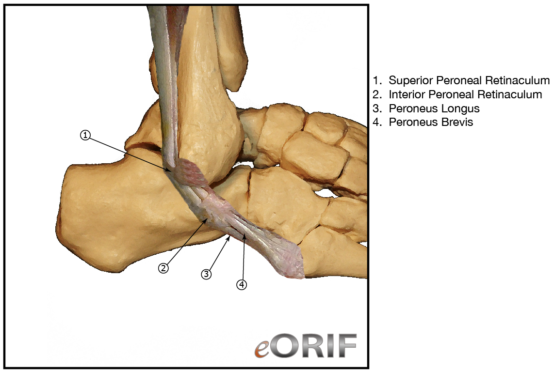
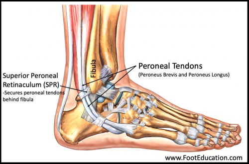
They are misdiagnosed as a lateral ankle sprain most of the time. The tendons are kept in place by the presence of a shallow groove on the posterior fibula the superior peroneal retinaculum spr which tethers the tendons to the fibula and a fibrocatilaginous lip which deepens the groove where it meets the retinaculum. Peroneal tendonitis peroneal subluxation and peroneal tendon tears.
Space compartment peroneal tendons contained within a common synovial sheath that splits at the level of the peroneal tubercle.
 Peroneal Tendinosis Footcaremd
 Peroneal Tendon Injuries Trinity Foot And Ankle Doctors
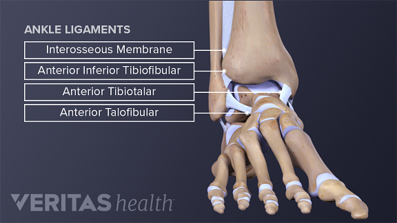 Ankle Anatomy Muscles And Ligaments
 Peroneal Tendonitis Tendinopathy Symptoms Causes
 Anatomy Of The Peroneus Longus Muscle Everything You Need To Know Dr Nabil Ebraheim
 The Fasciae Around The Ankle Human Anatomy
 Accessory Peroneal Muscles Radiology Reference Article
 Doc On The Run Podcast Peroneal Subluxation In Runners
 Peroneal Tendon Rupture
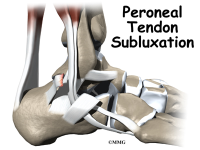
 Peroneal Tendon Subluxation
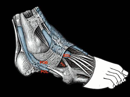
 Lateral View Of The Foot And Ankle Showing The Commonly
 Summit Medical Group
 Peroneal Tendon Subluxation Dislocation Foot Ankle
 Custom Made Insoles For Peroneal Tendonitis Boyner Clinic
 Peroneal Tendon Subluxation Eorthopod Com

 Peroneus Brevis Tendon Strain Symptoms Causes Treatment
 Posterior Tibial Tendon Insufficiency Ptti Foot Ankle
 Ankle Foot Anatomy
 Peroneal Tendon Syndromes Practice Essentials Epidemiology
 Understanding Subtle Peroneal Subluxation Treating The
 Tendons And Muscles Of The Foot And Ankle Including The Bones
 0086 2015 05 22 Bluman Peroneal Mp4

 Peroneal Tendon Subluxation Dislocation Foot Ankle
 Acute Peroneal Tendon Subluxation Footeducation
 Peroneal Tendon Dislocation S86 399a 726 79 Eorif
 Peroneal Tendonitis Part 1 Ankle Anatomy Foot Anatomy
 Chronic Peroneal Tendon Subluxation Footeducation
Unusual Accessory Peroneal Muscles Peroneus Quartus
Belum ada Komentar untuk "Peroneal Tendon Anatomy"
Posting Komentar