Sectional Anatomy Of Spinal Cord
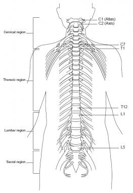
Anatomy of the spinal cord. The cervical thoracic lumbar and sacral nerves.
 Cross Sectional Anatomy The Central Nervous System
The two grooves are named as follows.
Sectional anatomy of spinal cord. At every segment there is a pair of right and left spinal nerves. Surrounding the canal is a single layer of cells the ependymal layer. Two prominent grooves or sulci run along its length.
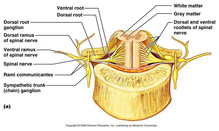
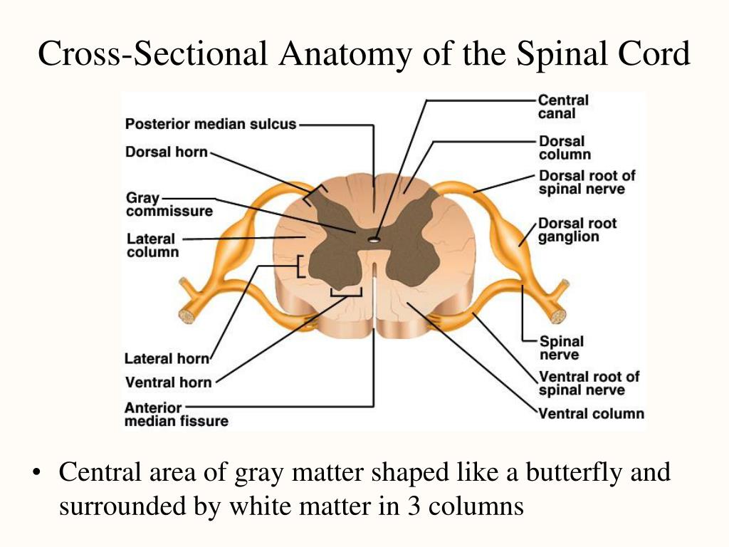
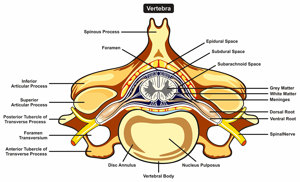
The spinal cord is part of the central nervous system cns which extends caudally and is protected by the bony structures of the vertebral column. The posterior median sulcus is the groove in the dorsal side and the anterior median fissure is the groove in the ventral side. An interactive quiz covering spinal cord cross sectional anatomy through multiple choice questions and featuring the iconic gbs illustrations.
The central gray matter contains the neural cell bodies. From each of these 6 to 8 nerve rootlets branch out in a definite and regular pattern. It contains the somas dendrites and proximal parts of the axons of neurons.
The spinal cord is elliptical in cross section being compressed dorsolaterally. Gray matter has a relatively dull color because it contains little myelin. It is covered by the three membranes of the cns ie the dura mater arachnoid and the innermost pia mater.
The ventral anterior median fissure and the more shallow dorsal posterior median sulcus. The spinal cord like the brain consists of two kinds of nervous tissue called gray and white matter. The gray matte of the spinal cord is located at its center with the white matter surrounding it.
Cross sectional anatomy of the spinal cord. Describe the cross sectional anatomy of the spinal cord and the location and function of the major spinal cord tracts. The spinal cord itself extends from the foramen magnum all the way to the first or second lumbar vertebra being enclosed inside the vertebral column.
These two grooves run the length of the cord and partially divide it into right and left halves. A cross sectional view of the spinal cord demonstrates a central butterfly shaped area of gray matter and peripheral white matter fig. The spinal cord is divided into four major parts.
The spinal cord medulla and the brain together make up the central nervous system cns. Anatomy and physiology of the spinal cord. A transverse section of the adult spinal cord shows white matter in the periphery gray matter inside and a tiny central canal filled with csf at its center.
Cross sectional anatomy of spinal cord. Collectively the entire spinal cord is divided into 31 segments. 34 internal structure of the spinal cord.
 Anatomy Of The Human Brain Prezentaciya Onlajn
 Nerve Wikipedia
Topographic And Functional Anatomy Of The Spinal Cord Gross
 Cross Sectional Anatomy An Overview Sciencedirect Topics
 Anatomy Central Nervous System Crosssectional Anatomy Stock
 Spinal Cord Anatomy Structure Function Tracts
 Spinal Cord And Autonomic Ns
 Spinal Cord Anatomy And Physiology I
 Neuraxial Anatomy Nysora
 Cross Sectional Anatomy Of The Spinal Cord A
 Cross Section Of Spinal Cord 1 Anatomy
 General Cross Sectional Anatomy Of The Spinal Cord
 Functions Of The Spinal Cord
 Ppt Chapter 14 The Central Nervous System Powerpoint
 Topographic And Functional Anatomy Of The Spinal Cord Gross
 Duke Neurosciences Lab 2 Spinal Cord Brainstem Surface
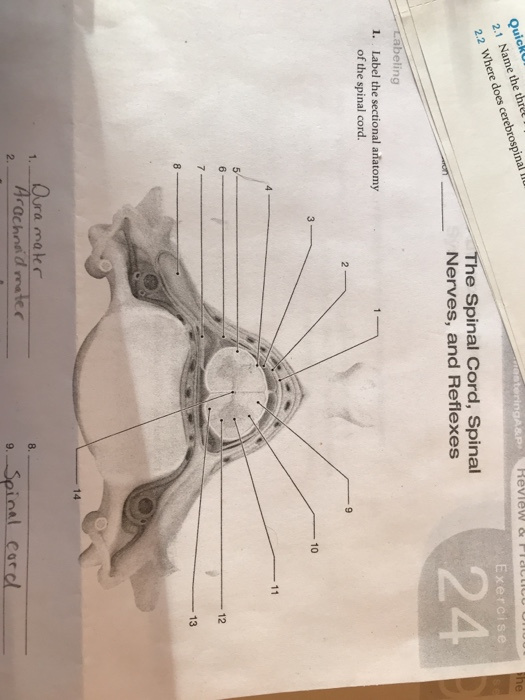 Solved Qu 2 Exercise 24 2 Where The Spinal Cord Spinal N
 Cross Section Of The Spinal Cord Anatomy At University Of
 Spinal Cord And Autonomic Ns
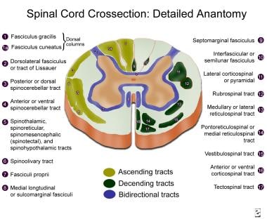
 Spinal Cord Sectional Anatomy In Detail
 Cross Sectional Anatomy Of The Spinal Cord Diagram Quizlet
 Applied Cross Sectional Anatomy Of Spinal Cord
 Spinal Cord Injuries Non Traumatic Nursing Ceu Aota
 Applied Cross Sectional Anatomy Of Spinal Cord
 Nervous System Anatomy Cross Section Anatomy Spinal Cord
 Functional Anatomy Of The Spinal Cord Springerlink
 Spinal Cord And Autonomic Ns


Belum ada Komentar untuk "Sectional Anatomy Of Spinal Cord"
Posting Komentar