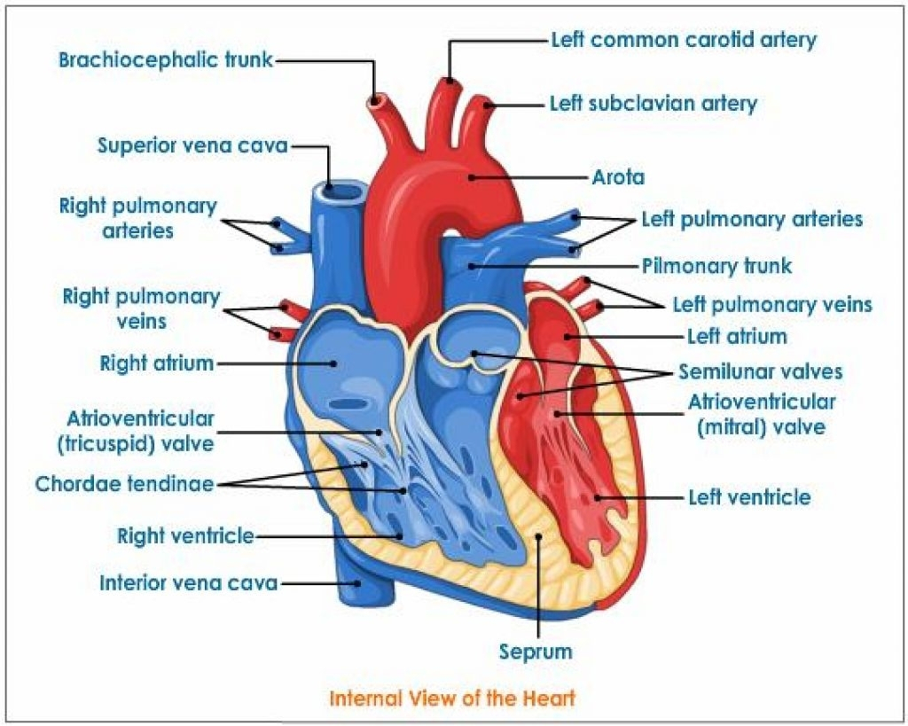
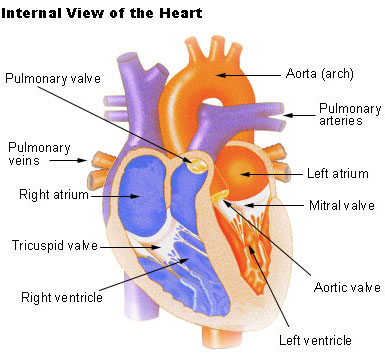
Internal Heart Anatomy
They form a figure 8 pattern around the atria and around the bases of the great vessels. It is the contraction of the myocardium that pumps blood through the heart and into the major arteries.
 3 D Printed Internal Heart Structures Model Amazon Com
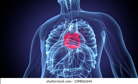
The heart pumps blood through the network of arteries and veins called the cardiovascular system.

Internal heart anatomy. Because the heart points to the left about 23 of the hearts mass is found on the left side of the body and the other 13 is on the right. Heart anatomy external receives blood from the left atrium via the mitral valve during diastole and is responsible for moving oxygenated blood to the systemic vasculature and organs during systole ventricular contraction. The muscle pattern is elegant and complex as the muscle cells swirl and spiral around the chambers of the heart.
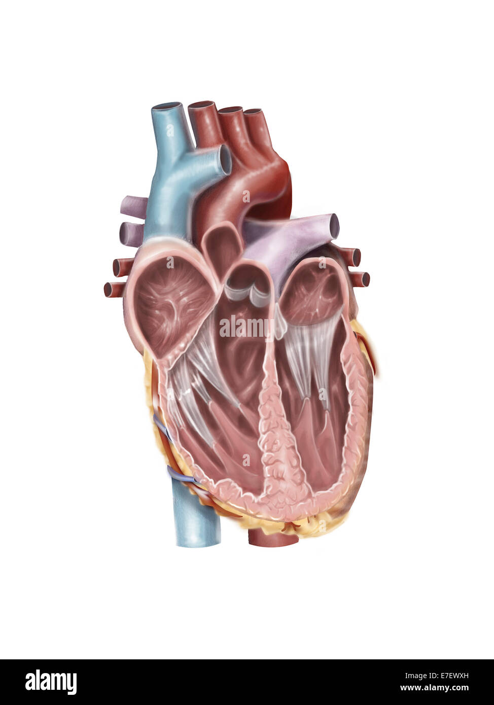
Internal anatomy of the heart study guide by christinostlund includes 41 questions covering vocabulary terms and more. Anatomy of the human heart internal structures. The heart ventricular walls consist of three layers.
Just pick an audience or yourself and itll end up in their incoming play queue. 0 0000 a shoutout is a way of letting people know of a game you want them to play. The anatomy of the heart.
Quizlet flashcards activities and games help you improve your grades. The heart is situated within the chest cavity and surrounded by a fluid filled sac called the pericardium. The 1epicardium the 2myocardium cardiac muscle and the skip to primary navigation skip to content.
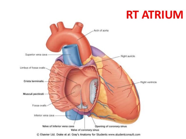
The right atrium receives blood from the veins and pumps it to the right ventricle. The heart has four chambers. Considered a higher pressure system compared to the right ventricle.
The right ventricle receives blood from the right atrium and pumps it to the lungs where it is loaded with oxygen. The walls and lining of the pericardial cavity are a special membrane known as the pericardium. The heart sits within a fluid filled cavity called the pericardial cavity.
This amazing muscle produces electrical impulses that cause the heart to contract. Anatomy of the heart pericardium. It is divided by a partition or septum into two halves and the halves are in turn divided into four chambers.
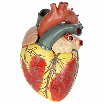 Amazon Com Alkita Human Heart Anatomy Model 3 Times Life
 Heart Anatomy Internal Heart Anatomy Cardiac Anatomy
 The Human Heart External And Internal Structure Online
 Heart Anatomy Chambers Valves And Vessels Anatomy

 The Heart
![]() Heart Right And Left Atrium Anatomy And Function Kenhub
Heart Right And Left Atrium Anatomy And Function Kenhub
![]() Heart Anatomy Structure Valves Coronary Vessels Kenhub
Heart Anatomy Structure Valves Coronary Vessels Kenhub
 3 Internal Features Of The Heart
![]() Human Internal Organs By Svitlana Babych
Human Internal Organs By Svitlana Babych
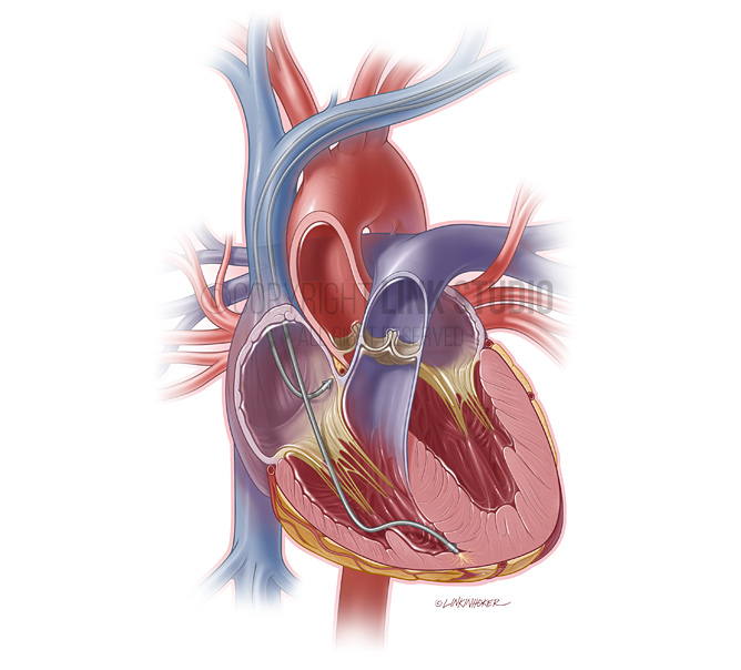 Heart Anatomy Link Studio
![]() Heart Anatomy Structure Valves Coronary Vessels Kenhub
Heart Anatomy Structure Valves Coronary Vessels Kenhub

 Practical 3 Internal External Heart Anatomy Physiology
 Internal Structure Of Heart Heart Diagram Heart
 3 Internal Structure Of The Heart
 Human Heart Diagram And Anatomy Of The Heart Heart
Heart Anatomy Isometric Isolated Stock Vector Colourbox
 Seer Training Structure Of The Heart
 Heart C Internal Anatomy
 Internal Heart Anatomy Part 2 Diagram Quizlet
 Free Anatomy Quiz Anatomy Of The Heart Quiz 1
![]() Heart Ventricles Anatomy Function And Clinical Aspects
Heart Ventricles Anatomy Function And Clinical Aspects
 Imagenes Fotos De Stock Y Vectores Sobre Internal Heart
 Internal Heart Anatomy Anterior Diagram Quizlet
 19 1 Heart Anatomy Anatomy And Physiology
 Close Up Of Internal Organs Dummy On White Background Human
 Internal View Of The Human Heart Stock Photo 73471737 Alamy

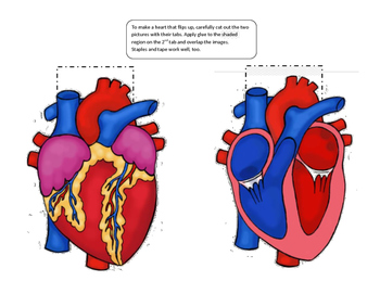 Paper Models Internal External Anatomy Of The Heart Booklet Foldable
Belum ada Komentar untuk "Internal Heart Anatomy"
Posting Komentar