Bronchial Anatomy
Each lung consists of. If desired labels on the map can be added or removed by clicking on the labeling controls below the map.
 Research Topics In Basic Sciences Anatomy Dr S Venkatesan Md
The trachea divides at the carina forming the left and right main stem bronchi which enter.

Bronchial anatomy. Location structure and anatomy the bronchi are located in the thoracic cavity along with the trachea and lungs. Bronchial atresia is a rare congenital disorder that can have a varied appearance. The cilia present in the luminal aspect of epithelial cells are in charge of the rhythmic upward movement of bronchial secretions from within the lung to the pharynx.
The respiratory epithelial cells are composed of basal bodies. The left lung is subdivided into two lobes and thereby into eight. It originates from the lower end of the trachea or windpipe where it divides or bifurcates at the point of carina into the left and right bronchus.
Lobes two or three these are separated by fissures within the lung. This allows the user to test their knowledge. Each lung has a tube called a bronchus that connects to.
The lungs are pyramid shaped paired organs that are connected to the trachea by the right and left bronchi. An infection of the lungs large airways bronchi usually caused by a virus. A bronchus which is also known as a main or primary bronchus.
On the inferior surface the lungs are bordered by the diaphragm. Apex the blunt superior end of the lung. The presence of mucin water and electrolytes contributes to the solubility of bronchial secretions.
Surfaces three these correspond to the area of the thorax that they face. A form of interstitial lung disease. Zooming in on the map can be done by adjusting.
Bronchopulmonary segmental anatomy gross anatomy. A bronchial atresia is a defect in the development of the bronchi affecting one or more bronchi usually segmental bronchi and sometimes lobar. Cough is the main symptom of acute bronchitis.
The user can follow the path of the bronchoscope on the bronchial tree map. Bronchial tree navigation map. The primary bronchi have cartilage and a mucous membrane that are similar.
No gas exchange takes place in the bronchi. Right main primary bronchus. The defect takes the form of a blind ended bronchus.
The trachea is a tube that carries the air in and out of your lungs. The right lung is subdivided into three lobes with ten segments. The interstitium walls between air sacs become scarred making the lungs stiff and causing shortness of breath.
Base the inferior surface of the lung which sits on the diaphragm. Gross anatomy of the lungs. The diaphragm is the flat dome shaped muscle located at the base of the lungs and thoracic cavity.
Bronchial tree the lungs begin at the bottom of your trachea windpipe.
 Endoscopic Bronchial Anatomy In The Cat Sciencedirect
 Drawing Of The Bronchial Anatomy Recorded During The Second
 Bronchi Anatomy Function Definition
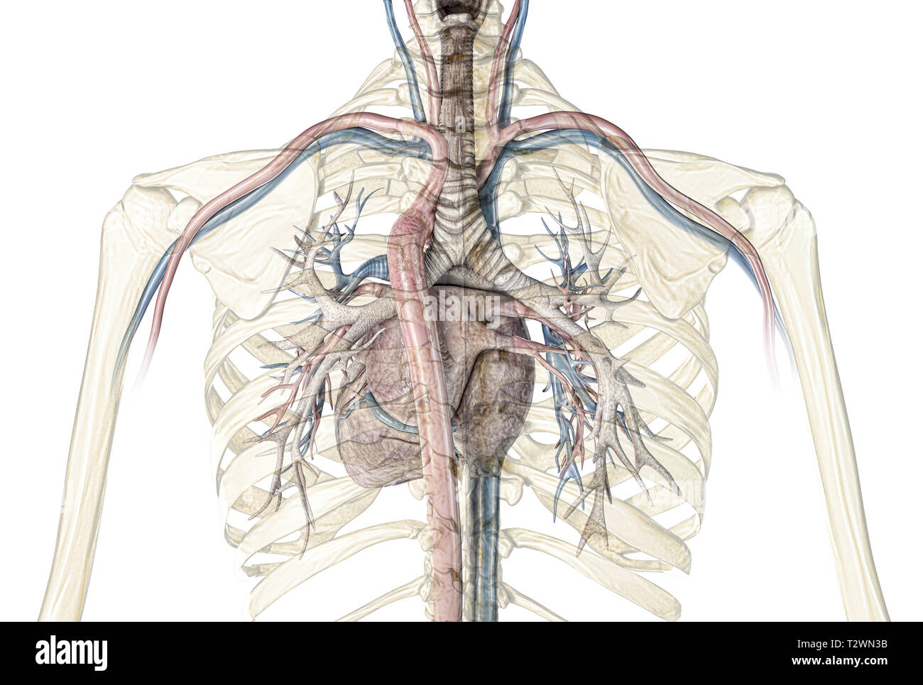 Human Heart With Vessels And Bronchial Tree Back View In
The Four Most Prevalent Patterns Of Bronchial Artery Anatomy
 Bronchopulmonary Segmental Anatomy Radiology Reference
 Anatomy Of The Trachea Carina And Bronchi Sciencedirect
 Four Main Types Of Bronchial Artery Anatomy Type I One
 Anatomy Of The Bronchus And Bronchial Tubes Poster
 Anatomy Lectures Thorax Bronchial Tree And Trachea
 Anatomy Of The Trachea Carina And Bronchi Sciencedirect
Pulmonary Circulation Anatomy And Peculiarities Humans
Bronchial Tree Architecture In Mammals Of Diverse Body Mass
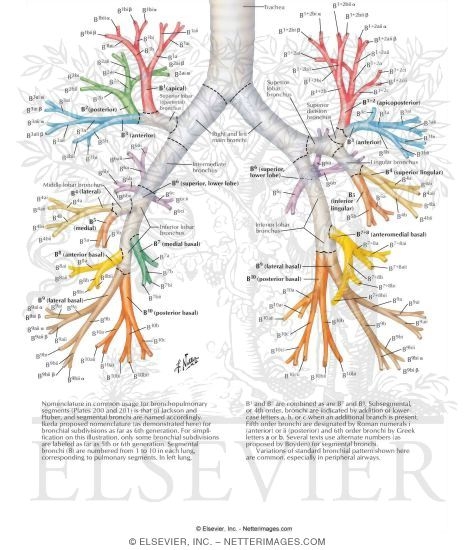 Nomenclature Of Bronchi Schema
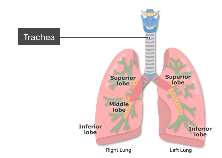 Bronchial Tubes Structure Functions Location Bronchus
 Magdy Said Anatomy Series Thorax 7 Thoracic Cavity Bronchial Tree Bronchopulmonary Segments V1
 Ppt Anatomy Of Tracheo Bronchial Tree Powerpoint
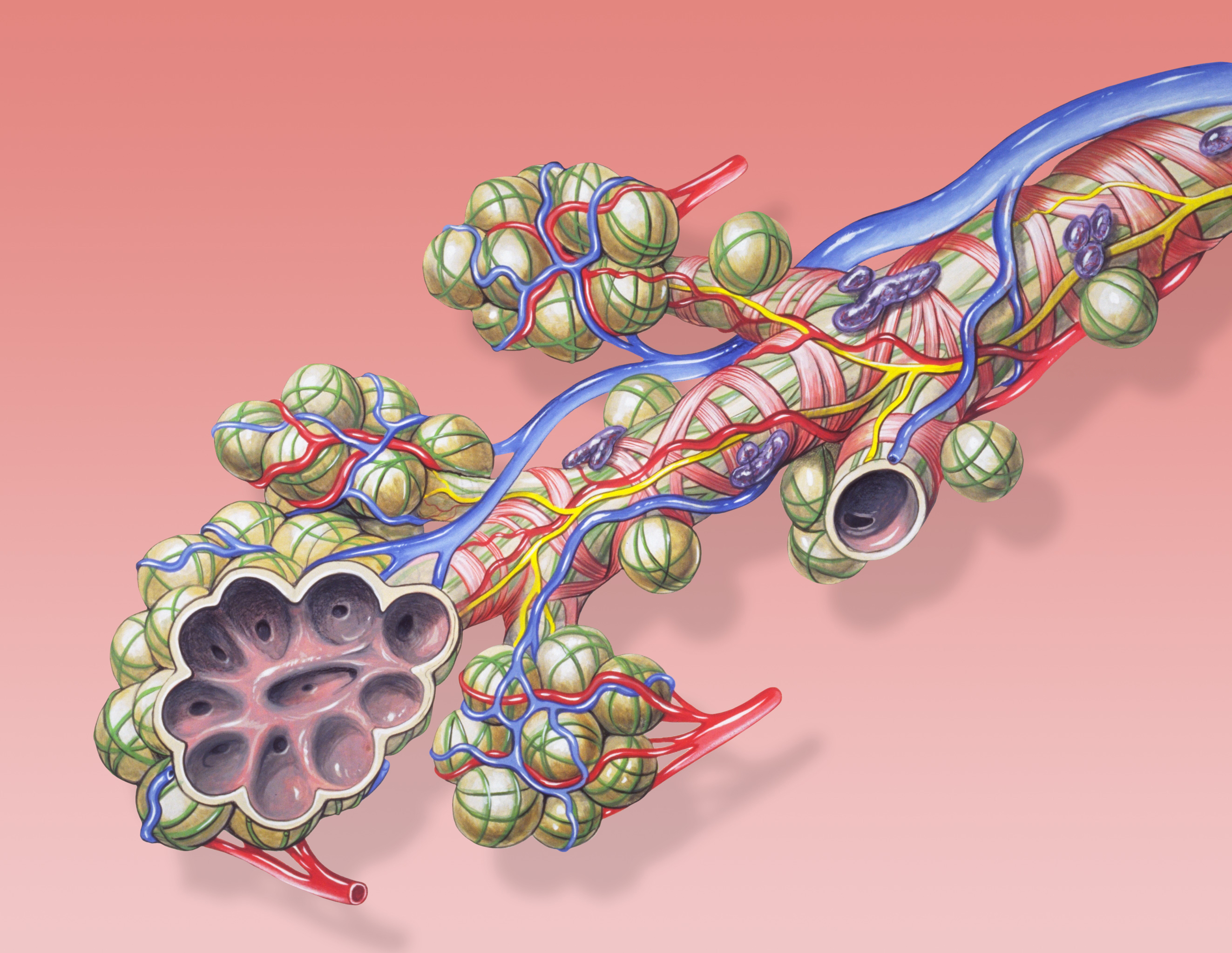 File Bronchial Anatomy Jpg Wikimedia Commons
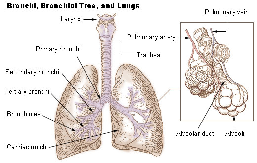 Seer Training Bronchi Bronchial Tree Lungs
 Lungs Origami Blue Paper Cut Human Lungs Anatomy With Bronchial


Belum ada Komentar untuk "Bronchial Anatomy"
Posting Komentar