Embryology Anatomy
Teachme anatomy part of the teachme series the medical information on this site is provided as an information resource only and is not to be used or relied on for any diagnostic or treatment purposes. It is however usually restricted to the phenomena which occur before birth.
 I Embryology 3 The Spermatozoon Gray Henry 1918
The term human anatomy comprises a consideration of the various structures which make up the human organism.

Embryology anatomy. After fertilisation there is the formation of the inner cell mass and outer cell mass and the zygote eventually becomes a blastocyst which is then ready for implantation. The embryonic structure or development of a particular organism. And λογία logia is the branch of biology that studies the prenatal development of gametes sex cells fertilization and development of embryos and fetuses.
It normally takes place in the ampullary region of the uterine tube. Additionally embryology encompasses the study of congenital disorders that occur before birth known as teratology. In a restricted sense it deals merely with the parts which form the fully developed individual and which can be rendered evident to the naked eye by various methods of dissection.
Human anatomy chapter 3 embryology. The term embryology in its widest sense is applied to the various changes which take place during the growth of an animal from the egg to the adult condition. Featuring a full color review of dental structures illustrated dental embryology histology and anatomy 4th edition provides a complete look at the development cellular makeup and morphology of the teeth and associated structures.
Schedule win gross anatomy unsw embryology awesome site simbryo animations. This is the process of male sperm fusing with the female ovum and its the basis of the embryology covered in the article. Fertilization is the process whereby genetic material from a male and female gamete fuse to form a single diploid nucleus.
A clear reader friendly writing style makes it easy to understand both basic science and clinical applications putting the material into the context of everyday dental practice. Embryology from greek ἔμβρυον embryon the unborn embryo. Fetus continues to grow and organs increase in complexity.
The remaining 30 weeks of development prior to birth when the organism is called a fetus. The branch of biology that deals with the formation early growth and development of living organisms. Embryology learning resources duke university medical school.
 Illustrated Dental Embryology Histology And Anatomy 5th
 Embryology Applied Anatomy And Physiology Of Lens
 Book Developmental Anatomy 1924 Embryology
 Bibliographie Anatomique Human Anatomy Histology

 Pdf Embryology Comparative Anatomy And Congenital
 Vagina Embryology Anatomy Histology Meduweb
 Atlas Of Neuroradiologic Embryology Anatomy And Variants
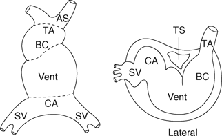 Cardiac Embryology And Anatomy Chapter 1 Core Topics In
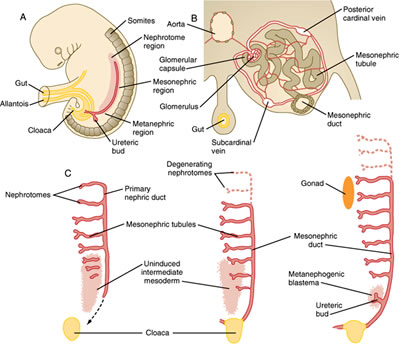 Duke Embryology Urogenital Development
Lecture Notes Lecture All Element 1 Anatomy Embryology
 Anatomy And Embryology Of The Eye 46 Slides Nice Review
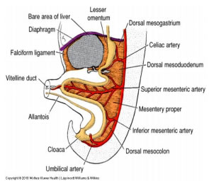 Duke Embryology Gut Development
 Embryology Anatomy And Histology Of The Kidney Springerlink
 The Brain And Cranial Nerves Chapter 9c The Brain
Veterinary Developmental Anatomy Courseware
Anatomy And Embryology Journals News And Media
 Embryology Of Normal Vascular Anatomy And Variant Celiac Sma
 Illustrated Dental Embryology Histology And Anatomy
Development And Anatomy Of The Pituitary Gland
 Evidence Of Evolution Embryological Expii
Veterinary Developmental Anatomy Courseware
 Anatomy And Embryology Of Rhodoscirpus Asper A Leaf Cross

 Embryology Anatomy And Diseases Of The Umbilicus Together With Diseases Of The Urachus Paperback
 Testis Development Embryology And Anatomy Springerlink
 Figure 1 From When Closure Fails What The Radiologist Needs
Annual International Conference Of Human Anatomy Embryology


Belum ada Komentar untuk "Embryology Anatomy"
Posting Komentar