Liver Lobes Anatomy
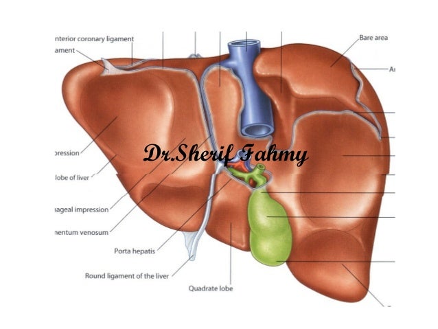
But the underside the visceral surface shows it to be divided into four lobes and includes the caudate and quadrate lobes. Each lobe is separated into many tiny hepatic lobules the livers functional units figure 3.
 Pediatric Liver Transplantation Practice Essentials Liver
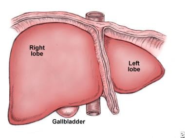
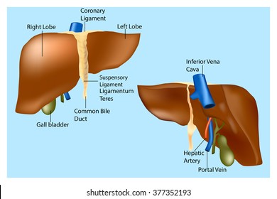
A fibrous capsule encloses the liver and ligaments divide the organ into a large right lobe and a smaller left lobe figure 2.

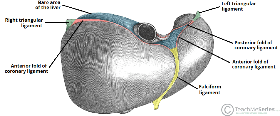
Liver lobes anatomy. The portal vein divides the liver into upper and lower segments. It is divided into a right lobe and left lobe by the attachment of the falciform ligament. Located on the lateral borders of the left and right lobes respectively the left and right triangular ligaments.
The falciform ligament runs inferiorly from the diaphragm across the anterior edge of. The liver is a vital organ found in humans and other vertebrates. There are two further accessory lobes that arise from the right lobe and are located on the visceral surface of liver.
The liver is grossly divided into two portions a right and a left lobe as viewed from the front diaphragmatic surface. The gallbladder sits under the liver along with parts of the pancreas and intestines. The liver also has two minor lobes the quadrate lobe and the caudate lobe.
It is a large organ with its major lobe occupying the right side of the abdomen below the diaphragm while the narrower left lobe extends all the way across the abdomen to the left. It lies between the inferior vena cava and a fossa produced by the ligamentum venosum a remnant of the fetal ductus venosus. Page needed the central vein joins to the hepatic vein to carry blood out from the liver.
Microscopically each liver lobe is seen to be made up of hepatic lobules. The wide coronary ligament connects the central superior portion of the liver to the diaphragm. The lobules are roughly hexagonal and consist of plates of hepatocytes radiating from a central vein.
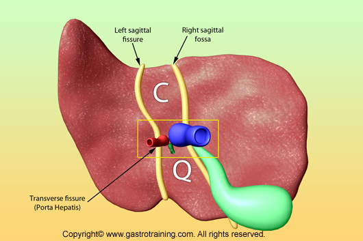
The falciform ligament visible on the front of the liver. Caudate lobe located on the upper aspect of the visceral surface. This plane runs from the inferior vena cava to the gallbladder fossa.
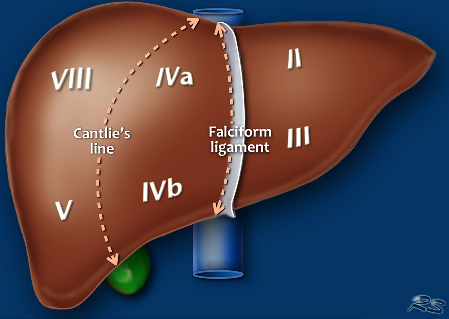
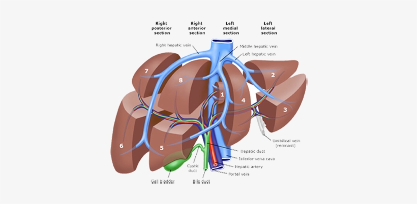
The liver has two large sections called the right and the left lobes. Middle hepatic vein divides the liver into right and left lobes or right and left hemiliver. The falciform ligament divides the left lobe into a medial segment iv and a lateral part segment ii and iii.

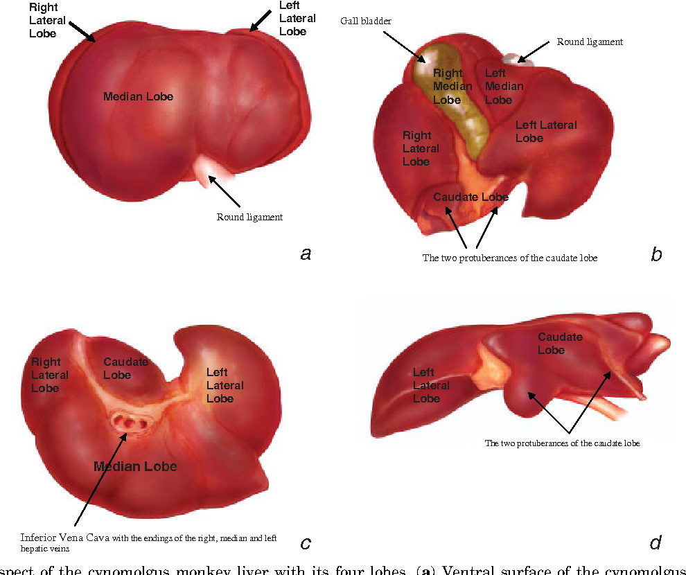 Figure 2 From First Description Of The Surgical Anatomy Of
![]() Human Liver Infographic Poster With Chart Diagram And Icon
Human Liver Infographic Poster With Chart Diagram And Icon
 The Liver Lobes Ligaments Vasculature Teachmeanatomy
 Liver Radiology Key
 The Liver Anatomy Of The Abdomen
 The Radiology Assistant Liver Segmental Anatomy
![]() Liver Functional Division Lobes And Segments Kenhub
Liver Functional Division Lobes And Segments Kenhub
 The Liver Lobes Ligaments Vasculature Teachmeanatomy
 Pld Liver Resection Polycystic Liver Disease Adpld Liver

![]() Liver Functional Division Lobes And Segments Kenhub
Liver Functional Division Lobes And Segments Kenhub
 Anatomy Of Cvs
![]() Liver And Gallbladder Anatomy Location And Functions Kenhub
Liver And Gallbladder Anatomy Location And Functions Kenhub
 Liver Biliary Anatomy Flashcards Quizlet
 Trauma Residents How To Remember Liver Anatomy The Trauma Pro

Liver Anatomy Ms1 Studocu
 Learning Objectives Liver Liver Hepatic Lobes Left Lobe
 Medical Textbook In The Net Liver Anatomy
Human Liver Anatomy Front Back And Two Lobes Location Of The
Liver Pancreas Spleen
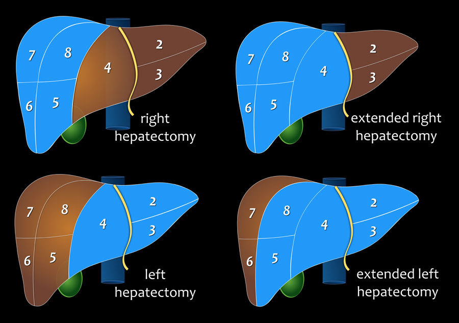 The Radiology Assistant Liver Segmental Anatomy

 Liver Lobe Images Stock Photos Vectors Shutterstock
 Porta Hepatis An Overview Sciencedirect Topics
 Imaging Essentials Small Animal Abdominal Ultrasonography

 Anatomical Liver With Gall Bladder Model
Liver Pancreas Spleen
 Porta Hepatis An Overview Sciencedirect Topics
 Liver Lobes Anatomy Liver Anatomy Bile Duct Dog Anatomy
Belum ada Komentar untuk "Liver Lobes Anatomy"
Posting Komentar