Pedicle Anatomy
Spine anatomy interactive video. The interpedicular distance was measured at the midshaft of the pedicle.

T12 usually has larger pedicle diameter than l1.

Pedicle anatomy. From a distance the spine often looks like a solid column but in fact it is made of individual vertebrae which are ring shaped bones that together form something. Connections between the left and the middle colic arteries are common. In articulates it develops from the caudal or hind region.
The vascular anatomy distal to the middle colic artery and near the splenic flexure is variable. In inarticulate larvae the pedicle a stalklike organ develops from a so called mantle fold along the valve margin. Glomerular filtration by slender cytoplasmic extensions called pedicels foot processes.
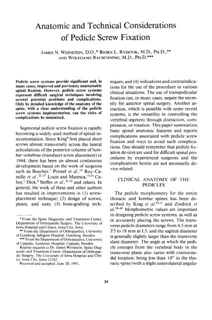
Biology a small stalk or stalklike structure especially one supporting or connecting an organ or other body part. Pedicle cervidae the attachment point for antlers in cervids. The most narrow dimensions in both the transverse and sagittal planes were chosen as the transverse pedicle width and the sagittal pedicle width respectively.
Most commonly 33 the ascending and descending branches of the left colic artery communicate through the marginal vessels. These processes are slightly expanded at their point of contact with the basement membrane and are separated from each other by slitlike spaces about 20 to 30 nanometres across. Pedicle diameter the pedicle wall is twice as thick medially as laterally.
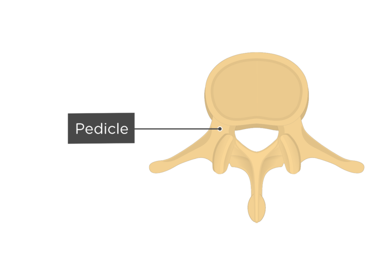
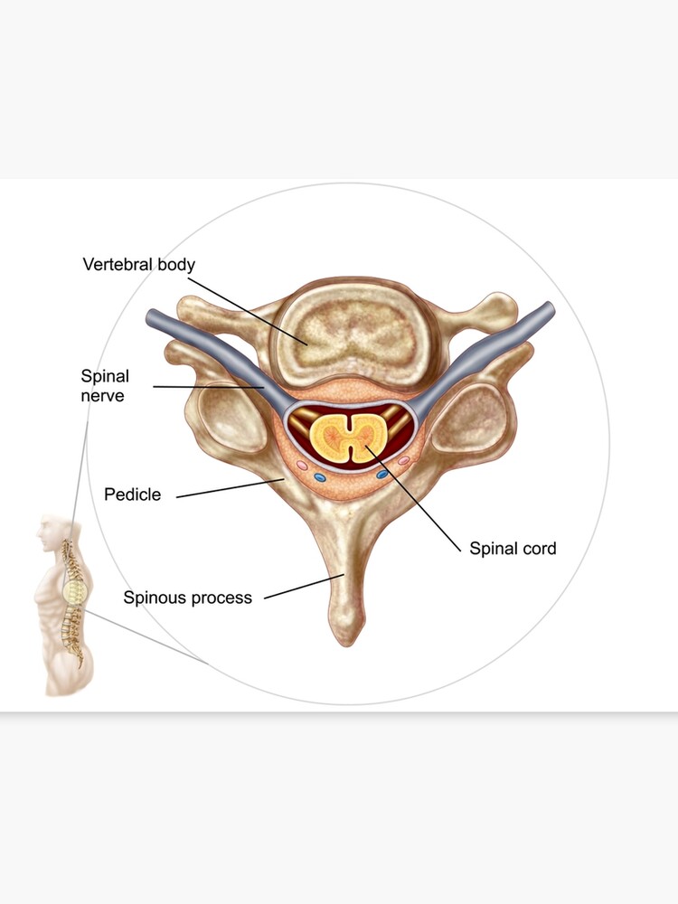
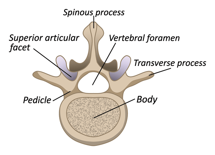
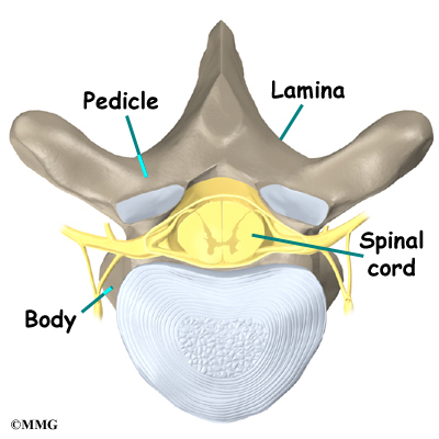
The pedicle is a stub of bone that connects the lamina to the vertebral body to form the vertebral arch. Also called pedicel. Two short stout processes extend from the sides of the vertebral body and joins with broad flat plates of bone laminae to form a hollow archway that protects the spinal cord.
Pedicel antenna the second segment of the antenna in the class insecta. The thoracic pedicle anatomy in all specimens was assessed by one researcher. Pedicel or petiole insect the stem formed by a restricted.
Strong cylindrical anatomic bridge between the dorsal spinal elements and the vertebral body. Animal anatomy pedicle in brachiopods a fleshy line used to attach and anchor brachiopods. Consists of a strong shell of cortical bone and a core of cancellous bone.
T4 has the narrowest pedicle diameter on average t7 can be irregular and have a narrow diameter on the concave side in ais. In anatomy the pedicle is a short protrusion on the inner part of each vertebra in the spine of all humans and many animals. It is important to remember that the pedicle size and angulation varies throughout the spinal column.
 Lumbar Vertebrae Anatomy
Anatomical And Technical Factors Associated With Superior
 General Osseous Anatomy Of The Spine Flashcards Quizlet
 Anatomic And Technical Considerations Of Pedicle Screw
 Innovative False Pedicle Surgery Allows For Advanced


 12180 01a T9 T10 Vertebrae Anatomy Exhibits
 Spine Anatomy About The Spine Virginia Spine Institute
 Lumbar Vertebrae An Overview Sciencedirect Topics
 Pedicle Identification Spine Surgery
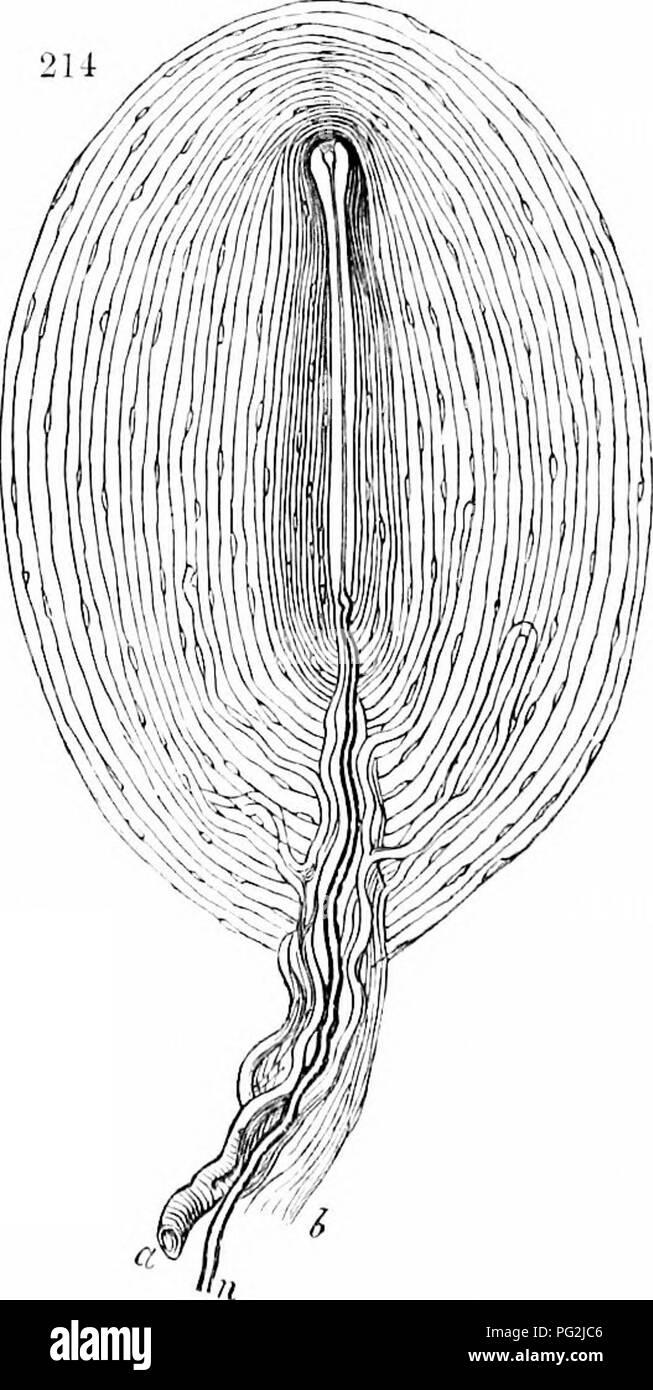
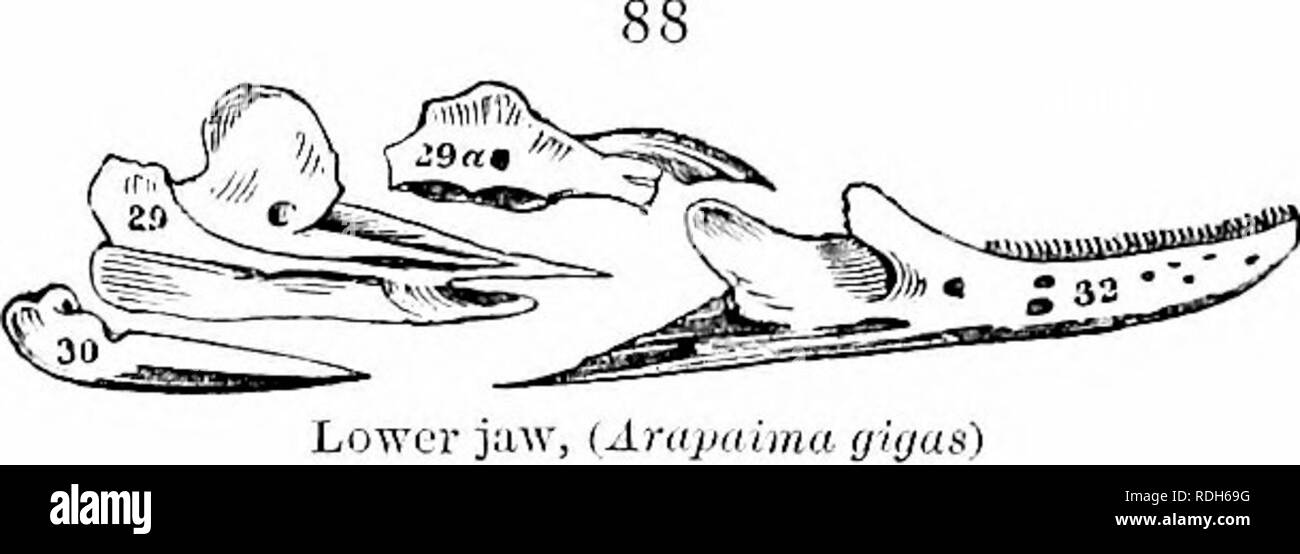 On The Anatomy Of Vertebrates Vertebrates Anatomy
 Anatomy Of Human Vertebra Canvas Print
 Anatomy Of The Spine Spinal Cord Injury Information Pages
 Ao Surgery Reference
 Anatomy Of The Vertebral Lumbar Column Vertebral Body
 Thoracic Vertebra An Overview Sciencedirect Topics
 Vertebra Wikipedia
 Nortex Spine Joint Institute Anatomy Of The Spine
 On The Anatomy Of Vertebrates Vertebrates Anatomy
 Dorsal Thoracic And Lumbar Screw Fixation And Pedicle
 Thoracic Spine Anatomy Orthogate
 Anatomy Atlases Illustrated Encyclopedia Of Human Anatomic

Belum ada Komentar untuk "Pedicle Anatomy"
Posting Komentar