Toe Bone Anatomy
The midfoot bones anatomy. Both conditions are treatable.
 Dorsal View Of The Bone Anatomy Of Rabbit Feet Left Front
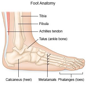
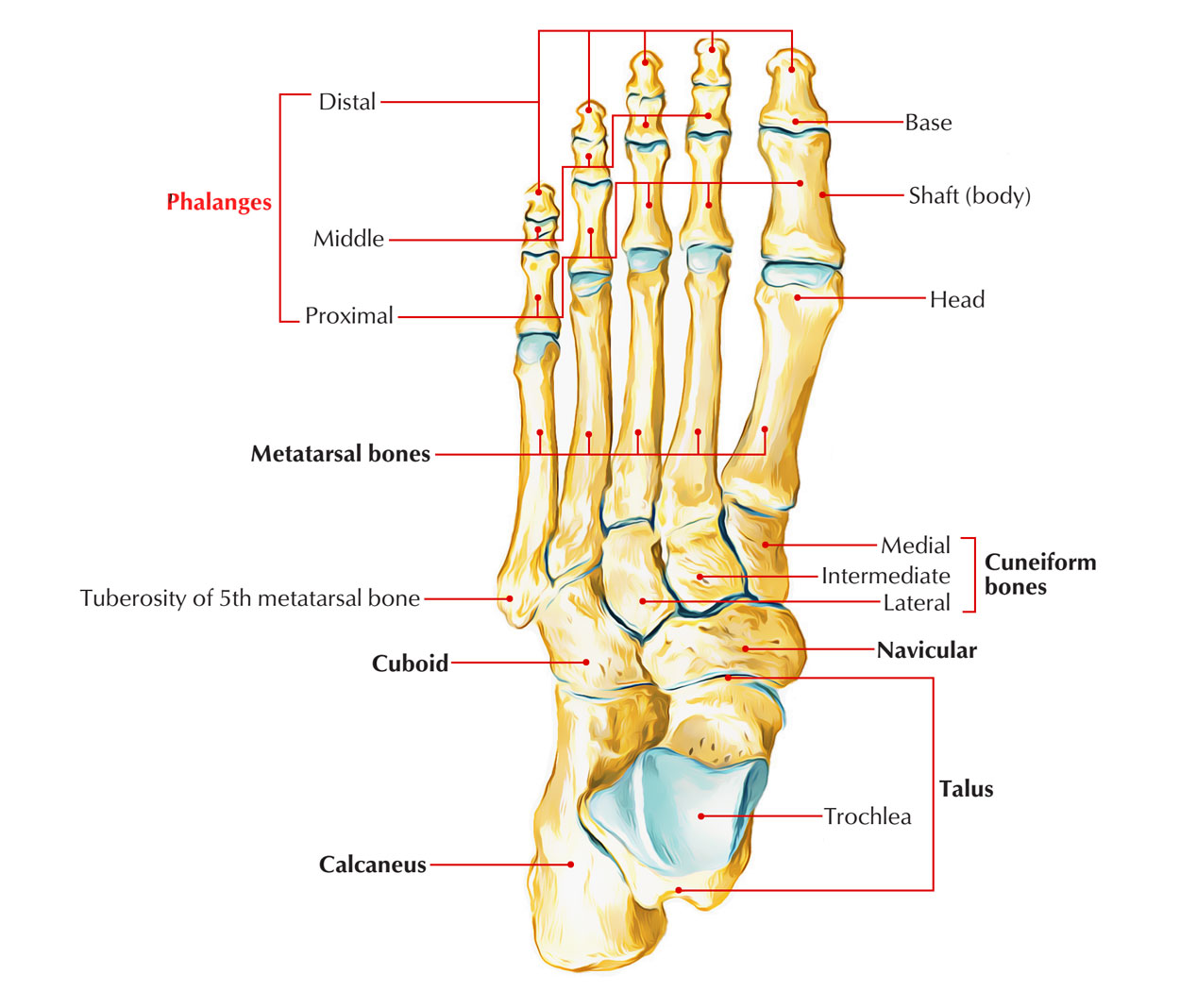
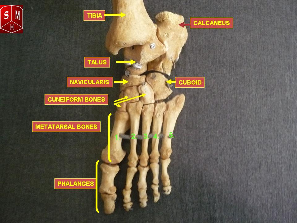
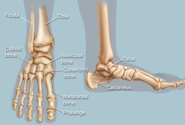
Talus the bone on top of the foot that forms a joint with the two bones of the lower leg.
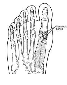
Toe bone anatomy. The midfoot bones are the navicular cuboid and cuneiform bones. Calcaneus the largest bone of the foot which lies beneath the talus to form the heel bone. The hindfoot forms the heel and ankle.
The bones of the feet are. A bunion is a progressive disorder that causes structural deformity of the bones and forefoot. Tarsals five irregularly shaped bones of the midfoot that form the foots arch.
This is an article covering the muscle attachments blood supply innervation and ossification of the phalanges of the foot. Toe movement is generally flexion and extension via muscular tendons that attach to the toes on the anterior and superior surfaces of the phalanx bones. In any normal foot you will find a total of 26 bones in each foot.
A bunion is a prominent bump on the inside of the foot near the base of the big toe. The metatarsals which run through the flat part of your foot. 28 bones if you count the 2 little sesamoid bones near the big toe joint.
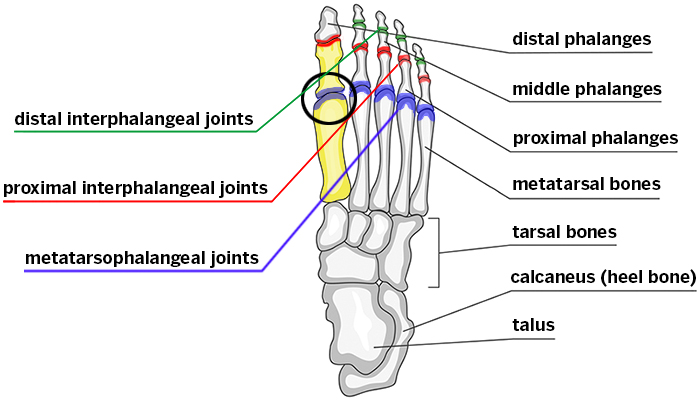
The navicular bone is one of the midfoot bones. The midfoot is made up of 5 tarsal bones. The phalanges which are the bones in your toes.
The feet are divided into three sections. The cuneiform bones the navicularis and the cuboid all of which function to give your foot. Together with the metatarsal bones proximal bones in the forefoot they form the arch of the foot.
Bones and main ligaments of the foot. The midfoot is a pyramid like collection of bones that form the arches of the feet. The talus which is the.
The calcaneus which is the bone in your heel. Learn this topic now at kenhub. Bunions develop when the bone at the base of the toe the first metatarsal begins to separate from the bone.
This in turn can cause the hallux to become misaligned from its normal position on the foot. Gout is caused by the deposit of uric acid crystals in the joint which results in periodic inflammation and pain. This is an article covering the articular surfaces ligaments and muscles that produce movement at the joints of the feet.
With the exception of the hallux toe movement is generally governed by action of the flexor digitorum brevis and extensor digitorum brevis muscles. Foot anatomy is quite detailed and more complex than most people think. The talus bone supports the leg bones.
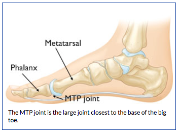
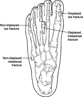
The forefoot contains the five toes phalanges and the five longer bones metatarsals. Each toe consists of three separate bones and two joints except for the big toe which has only two bones distal and proximal phalanges and one joint like the thumb in the hand.
Foot Anatomy Orthopedic Surgery Algonquin Il Barrington
 Toe Fracture In Children What You Need To Know
 Anatomy Of Turf Toe Bouldercentre For Orthopedics Spine
 Toe And Metatarsal Fractures Broken Toes Foot Health Facts
 The Newborn Foot American Family Physician
 Easy Notes On Skeleton Of The Foot Learn In Just 6
Human Being Anatomy Skeleton Foot Image Visual
 Bones Of The Foot Illustrations Foot Anatomy Illustrations
 The Foot Advanced Anatomy 2nd Ed
 Sesamoid Injuries In The Foot Foot Health Facts
Understanding Hallux Rigidus Limitus A Proven Toe Implant
 Hallux Rigidus Stiff Big Toe Hss Edu
 Foot Anatomy Bones Ligaments Muscles Tendons Arches
 Left Foot Ankle Bone Anatomy Bone Anatomy Of Foot Anatomy
 The Bones In The Foot Inferior View Picture Illustrated
 Anatomy 101 Strengthen Your Big Toes To Build Stability
 Foot Bones Anatomy And Mnemonic Tarsals Metatarsals
 How To Draw Feet With Structure Foot Bone Anatomy
:background_color(FFFFFF):format(jpeg)/images/library/11041/anatomy-ankle-joint_english.jpg) Ankle And Foot Anatomy Bones Joints Muscles Kenhub
Ankle And Foot Anatomy Bones Joints Muscles Kenhub
 Understanding And Caring For Your Feet Breaking Muscle
 Feet Human Anatomy Bones Tendons Ligaments And More


Belum ada Komentar untuk "Toe Bone Anatomy"
Posting Komentar