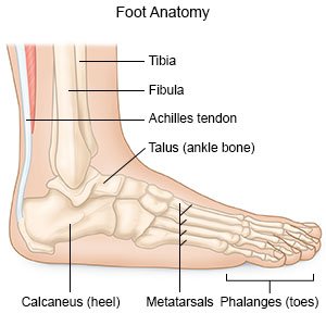
Anatomy Foot And Ankle
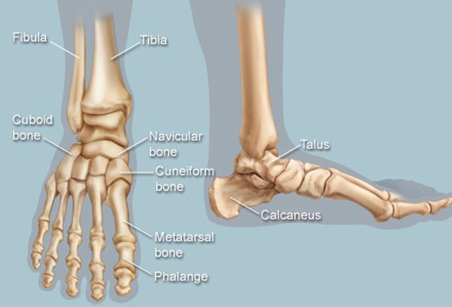
Upper ankle joint tibiotarsal talocalcaneonavicular and subtalar joints. The calcaneus heel bone is the largest bone in the foot.
 Ankle Foot Atlas Of Anatomy
The subtalar joint sits below the ankle joint and allows side to side motion of the foot.

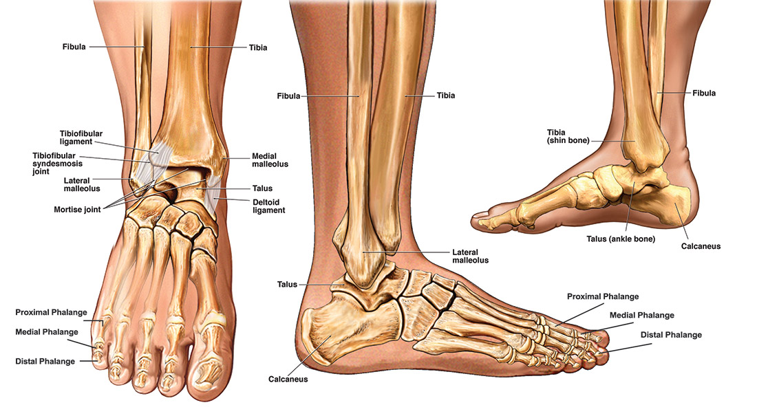
Anatomy foot and ankle. Together they form a bracket shaped socket covered in hyaline cartilage. The talus bone supports the leg bones tibia and fibula forming the ankle. It is made up of three joints.
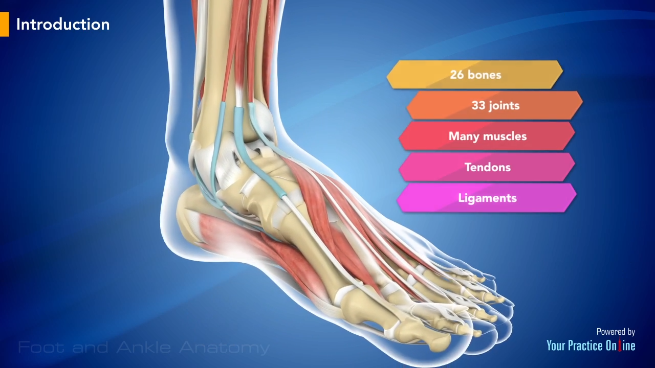
Sarrafians anatomy of the foot and ankle. 50 out of 5 stars 6. These all work together to bear weight allow movement and provide a stable base for us to stand and move on.
The most well known are the gastrocnemius and soleus muscles which are the strong calf muscles that allow you to push up on your toes. The ankle joint is formed by three bones. The strong muscles that move the ankle originate in the leg.
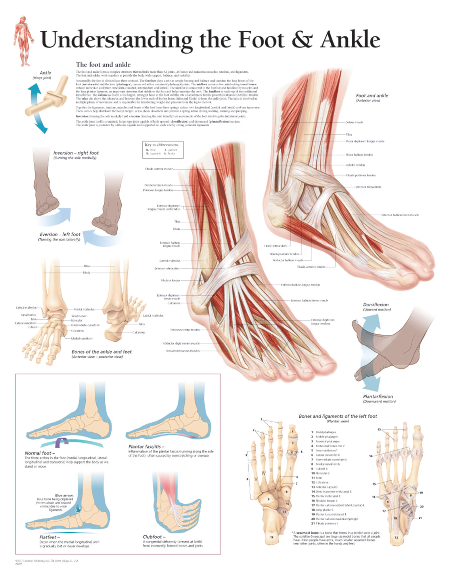
90 to rent 13970 to buy. The ankle joint allows up and down movement of the foot. The foot consists of thirty three bones twenty six joints and over a hundred muscles ligaments and tendons.
Free shipping by amazon. Ankle anatomy the ankle joint also known as the talocrural joint allows dorsiflexion and plantar flexion of the foot. In the lower leg are two bones called the tibia shin bone and the fibula.
In order to understand conditions that affect the foot and ankle it is important to understand the normal anatomy of the foot and ankle. Use our anatomy tools to learn about bones joints ligaments and muscles of the foot and ankle. By kelikian md armen s and sarrafian md facs shahan k.
The ankle consists of three bones attached by muscles tendons and ligaments that connect the foot to the leg. Foot and ankle anatomy is quite complex. Numerous ligaments made of tough moveable.
The tibia and fibula of the leg and the talus of the foot. Footeducation is committed to helping educate patients about foot and ankle conditions by providing high quality accurate and easy to understand information. Hardcover 9590 95.
Get it as soon as mon jul 29. The tibia and fibula are bound together by strong tibiofibular ligaments. There are many muscles that help to move and support the ankle and foot.
The hindfoot forms the heel and ankle. The last two together are called the lower ankle joint.
 Common Conditions Of The Foot And Ankle An Overview
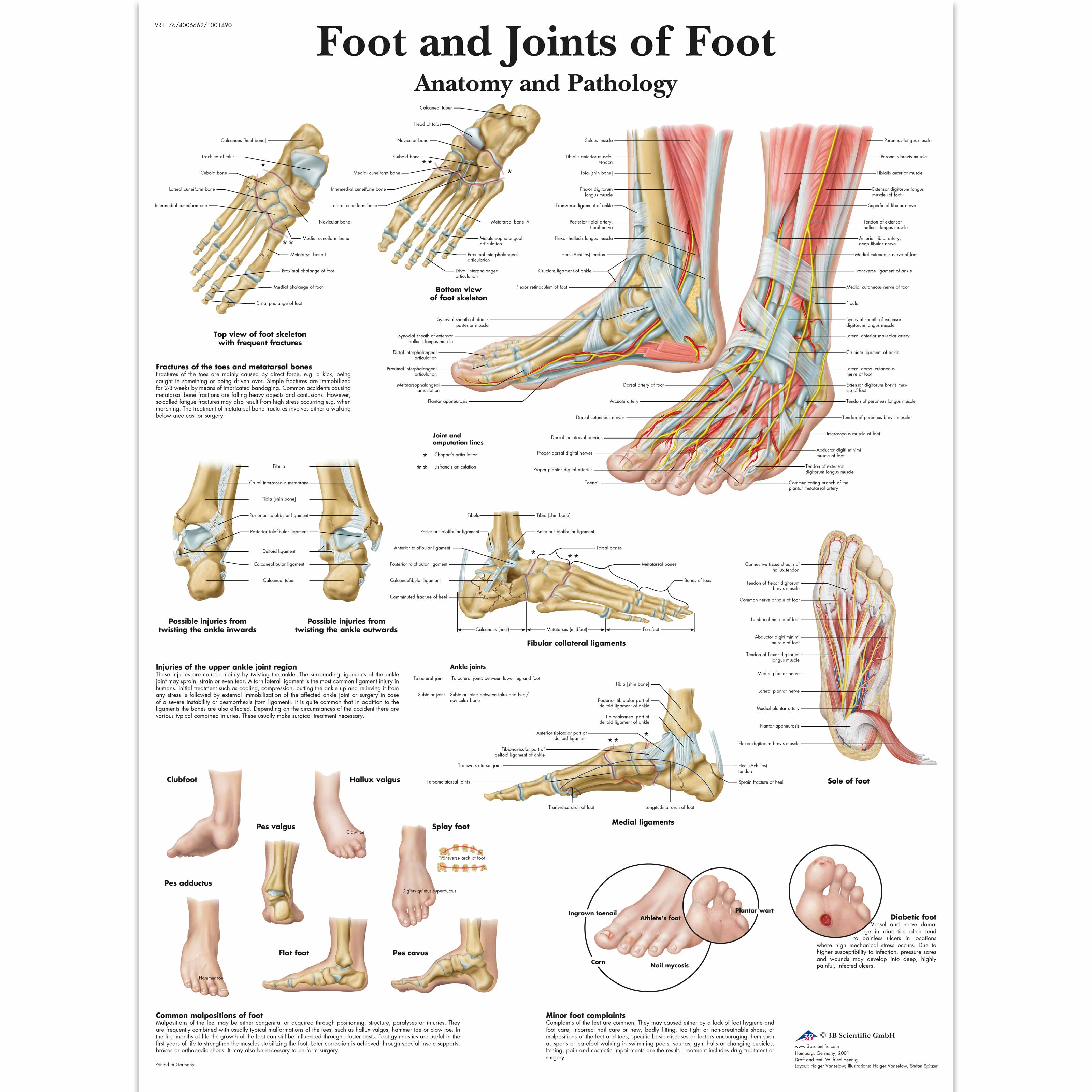
 Anatomy And Injuries Of The Foot And Ankle Chart 20x26 Clinicalposters
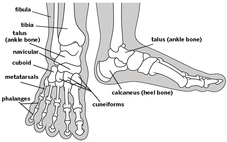
 Foot And Joints Of Foot Chart Anatomy And Pathology
 Applied Anatomy Of Ankle And Foot
 Bones The Of Foot Stock Vector Illustration Of Orthopedic
 Ankle Foot Anatomy
 Bones Of The Foot And Ankle Purposegames
 Ankle Fracture What You Need To Know
 Foot Bones Foot Pain Anatomy Info
 Foot And Ankle Anatomy Bones Muscles Ligaments Tendons
 Foot Ankle Foot Anatomy Ankle Anatomy Leg Anatomy

 Anatomy Of Foot And Ankle Poster Anatomical Chart Human Body Educational Home Decor
 12114 09x Normal Left Foot And Ankle Anatomy Exhibits

 Common Foot And Ankle Conditions Pro Sports Orthopedics
 Foot Anatomy Spokane Valley Wa Foot Doctor
 Feet Human Anatomy Bones Tendons Ligaments And More
 Axis Scientific Foot And Ankle Joint Section Anatomy Model
 Understanding The Foot Ankle
 Foot And Ankle Pain Podiatry St Joseph Amp 39 S
 Foot Bones Anatomy Conditions And More

 3b Scientific A31 1l Human Left Loose Foot And Ankle Skeleton
 Foot And Ankle Anatomy
 Understanding And Caring For Your Feet Breaking Muscle

 Anatomy And Injuries Of The Foot And Ankle Poster
 Ankle Sprains Summit Orthopedics
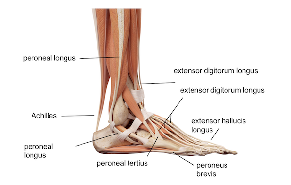 Ankle Foot Anatomy
 Ankle Foot Anatomy
Belum ada Komentar untuk "Anatomy Foot And Ankle"
Posting Komentar