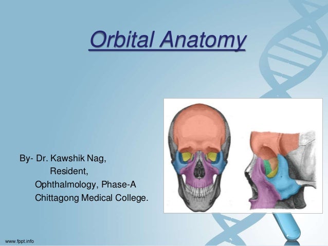
Orbital Anatomy
The orbital floor is the most frequently fractured wall in trauma where an object larger than the orbit such as a ball or fist impacts the entire orbit. Orbit can refer to the bony socket or it can also be used to imply the contents.
 Orbital Anatomy Tutorial
The childs orbit is rounder but with age the width increases.
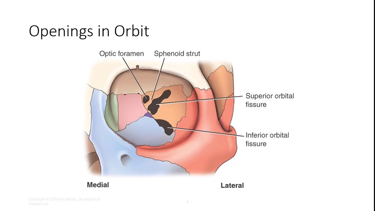
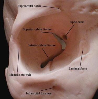
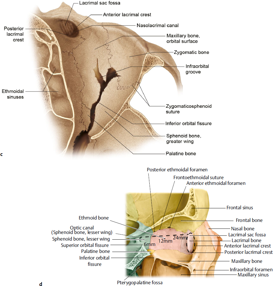
Orbital anatomy. Fig 12 the major openings into the orbit. The widest circumference of the orbit is inside the orbital rim at the lacrimal recess. The orbital floor is the only wall of the orbit that does not contain part of the sphenoid bone.
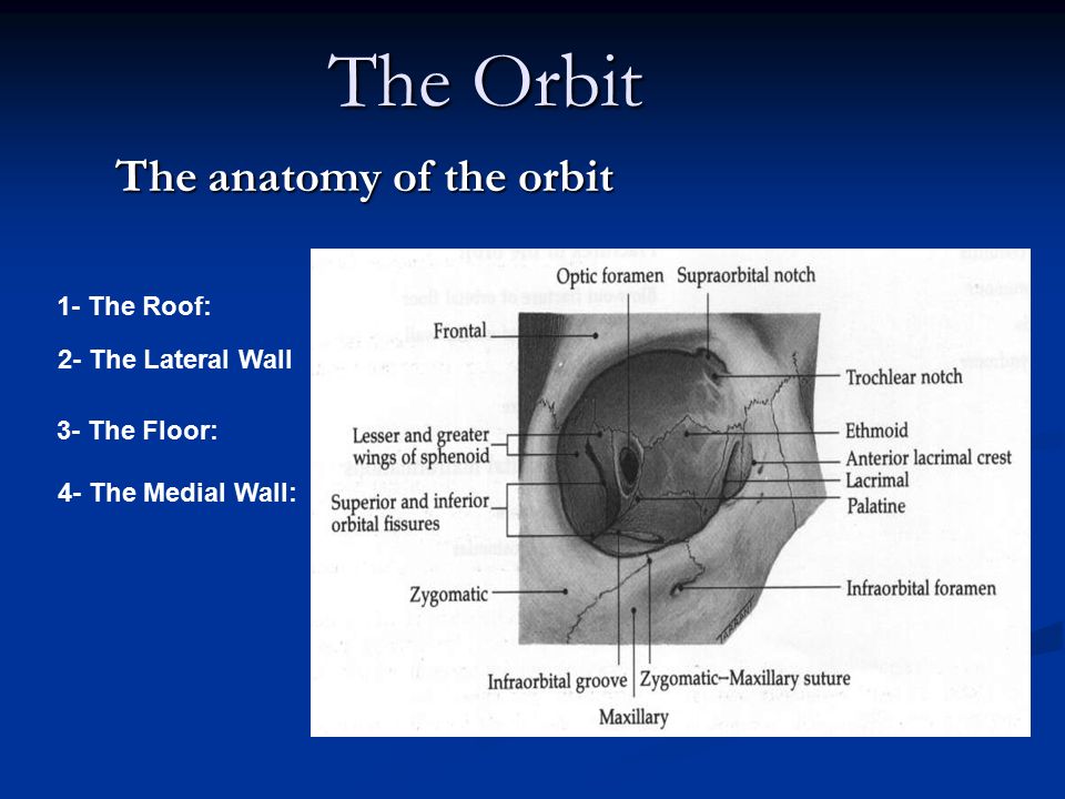
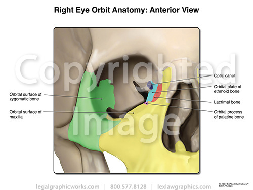
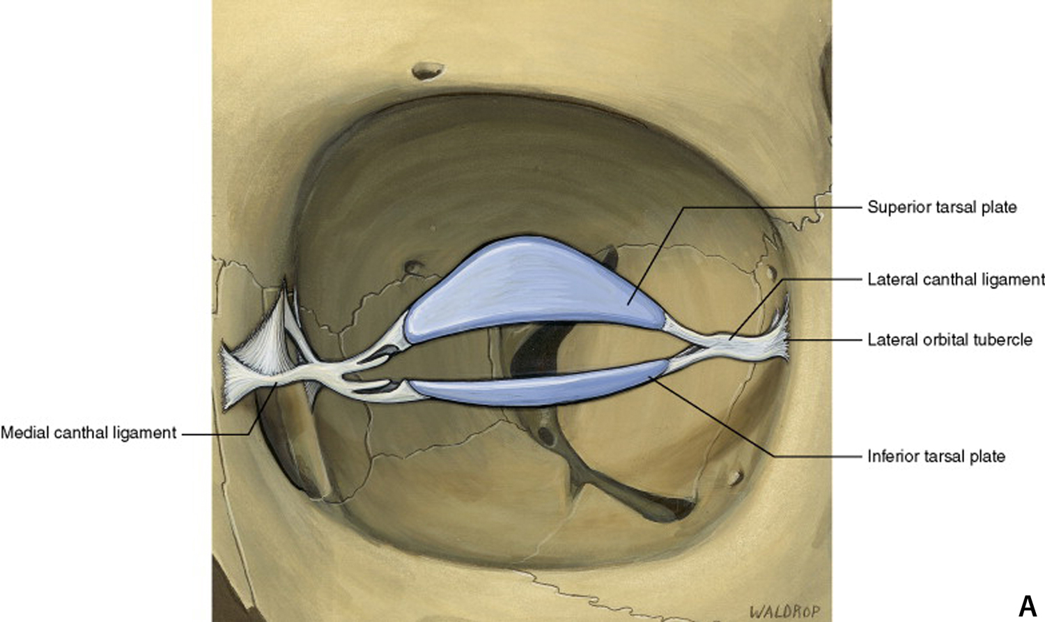
The bony orbit borders and anatomical relations. Orbital anatomy the orbital cavities are large bony sockets that house the eyeballs with associated muscles nerves blood vessels and fat. The contents of the orbit are separated and supported by multiple.
The orbit which protects supports and maximizes the function of the eye. Orbit anatomy orbit celestial mechanics orbit celestial mechanics orbit celestial mechanics orbit disambiguation orbit disambiguation orbit group theory orbit physics orbit physics orbit physics orbit adjust propulsion system. This fissure allows the passage to the nerves iii iv vi branches of the v1 and ophthalmic veins.
Orbital process of the frontal bone orbital process of the zygomatic bone. Use the mouse scroll wheel to move the images up and down alternatively use the tiny arrows on both side of the image to move the images. Fig 11 diagram of the arterial supply to the eye.
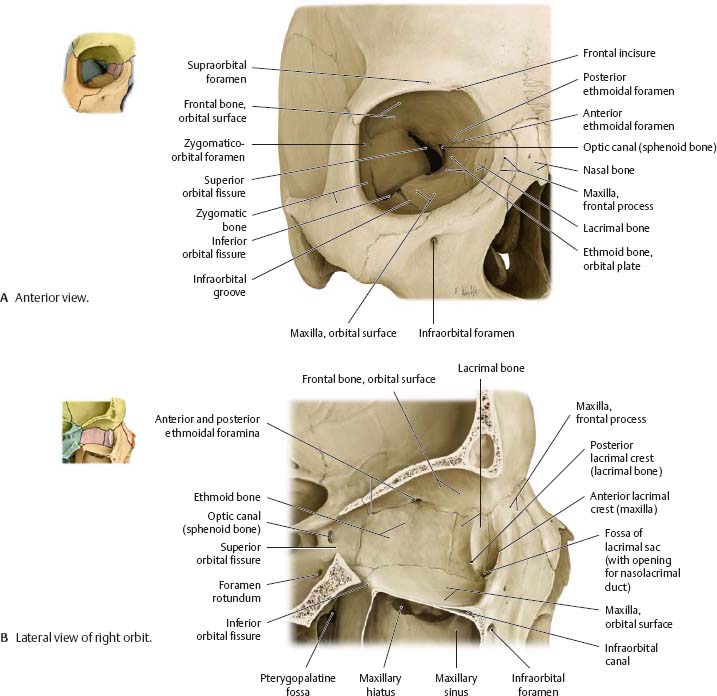
Inferior orbital fissure lies between. From the medial orbital rim to apex the orbit measures approximately 45 mm in length whereas from the lateral orbital rim to the apex the measurement is approximately 1 cm shorter. Pathways into the orbit.
In the adult human the volume of the orbit is 30 millilitres 106 imp fl oz. 101 us fl oz. Orbit anatomy in anatomy the orbit is the cavity or socket of the skull in which the eye and its appendages are situated.
Superior orbital fissure lies between the lesser and the greater wing of sphenoid. The lacrimal system produces distributes and drains tears. Orbit and attitude tracking.
The orbit can be thought of as a pyramidal structure. This mri orbits and paranasal sinuses cross sectional anatomy tool is absolutely free to use. Each orbit is pear shaped with the optic nerve representing the stem.
 Anatomy Of The Eye And Orbit The Clinical Essentials
 Orbital Anatomy And Its Clinical Applications Ento Key
 Orbital Floor Blowout Fracture Brown Emergency Medicine
 Superior Orbital Fissure Wikipedia
 Schematic Diagrams Of Orbital Anatomy A Schematic Diagram
 Orbital Tumor Eye Socket Cancer Anatomy
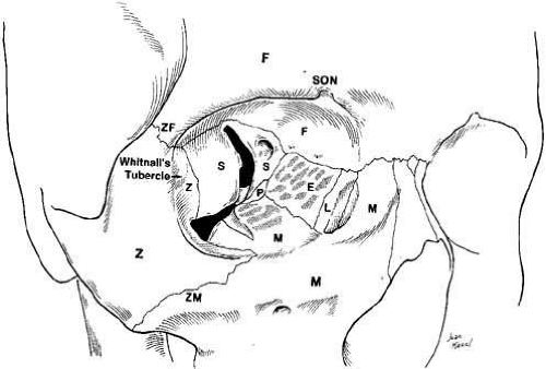
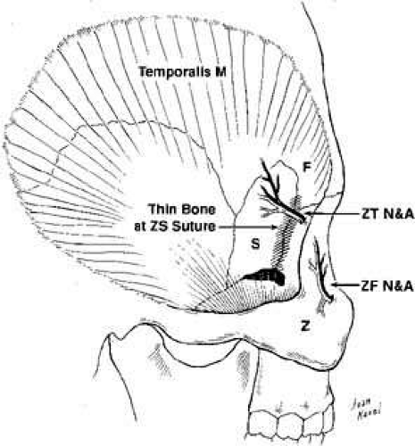
 Midface Authors Added Material Ao Surgery Reference
 Orbits And Eyes Anatomical Illustrations
![]() Bones Of The Orbit Anatomy Foramina Walls And Diagram
Bones Of The Orbit Anatomy Foramina Walls And Diagram
![]() Bones Of The Orbit Anatomy Foramina Walls And Diagram
Bones Of The Orbit Anatomy Foramina Walls And Diagram
Surgical Anatomy Of The Cavernous Sinus Superior Orbital
 Orbital Anatomy And Its Clinical Applications Ento Key
 Anatomy The Orbit Flashcards Quizlet
 Orbital Bone Anatomy Eye Anatomy Facial Anatomy
 The Anatomy Of The Orbit Ppt Download
 Anatomy And Pathology Of The Orbits

 Skull Orbit Anatomy
 Orbital Anatomy Illustration Radiology Case
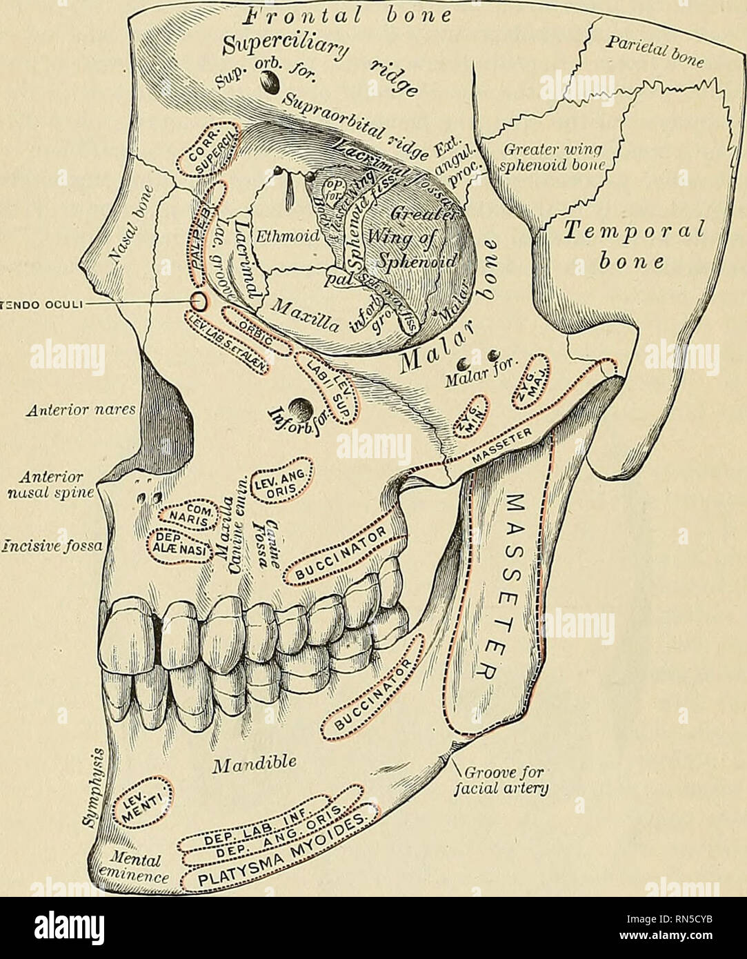 Anatomy Descriptive And Applied Anatomy 136 Special
 Orbit Eye Atlas Of Anatomy
 Orbit Anatomy Osteology Lacrimal System Connective Tissue
 Human Eye And Orbital Anatomy Superior View Illustration
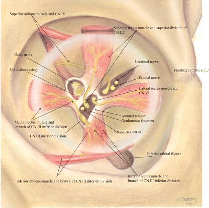
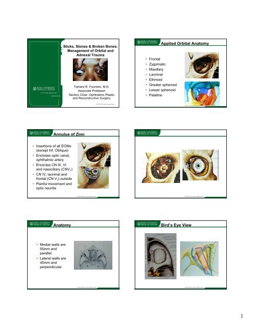 Applied Orbital Anatomy Annulus Of Zinn Anatomy Bird S Eye View
 Ocular Orbit
![]() Schematic Anatomy Of The Human Orbit In Transverse Cross
Schematic Anatomy Of The Human Orbit In Transverse Cross
![]() Eye Anatomy Muscles Arteries Nerves And Lacrimal Gland
Eye Anatomy Muscles Arteries Nerves And Lacrimal Gland
 Instant Anatomy Diagram
 Ppt Anatomy And Diseases Of The Orbit Powerpoint
 The Radiology Assistant Orbita Pathology
 Orbital Anatomy Plastic Surgery Key
 Schematic Diagrams Of Orbital Anatomy A Schematic Diagram
 Orbital Anatomy
Belum ada Komentar untuk "Orbital Anatomy"
Posting Komentar