Ct Sinuses Anatomy
For a ct scan of the sinuses the patient is most commonly positioned lying flat on the back. Axial view coronal view sagittal view.

To load the sinus ct anatomy module in a new window click on its image above.
Ct sinuses anatomy. Other functions are air humidification and aiding in voice resonance. Imaging the paranasal sinuses is routine in clinical practice to evaluate for various sinus pathology non specific facial pain and pre operative planning for functional endoscopic sinus surgery fess including post operative follow up. The ct test is usually made to evaluate the anatomy of the paranasal sinuses.
The module interface is meant to mimic a radiology workstation with adjacent image scrolling via arrow keys and or mouse wheel button. Straps and pillows may be used to help the patient maintain the correct position and to hold still during the exam. The patient may also be positioned face down with the chin elevated.
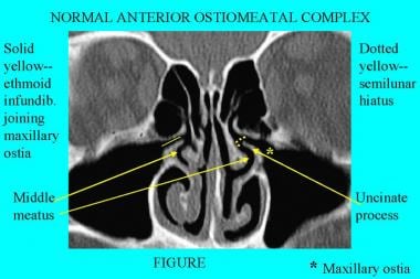
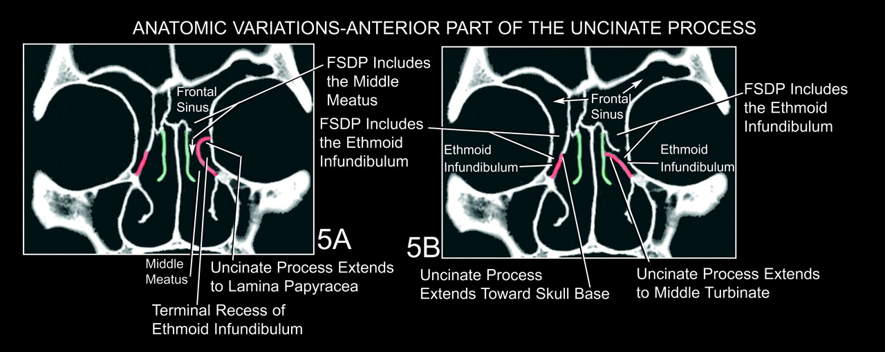
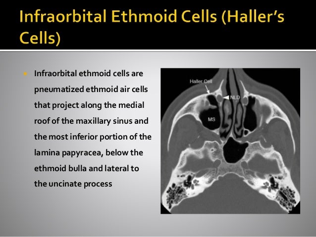
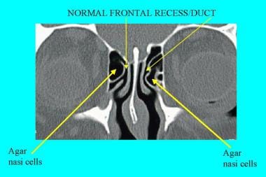
Its ability to optimally display bone soft tissue and air provides an accurate depiction of both the anatomy and the extent of disease in and around the paranasal sinuses. Numerous sinonasal anatomic variants exist and are frequently seen on sinus ct scans. The most common ones are agger nasi cells infraorbital ethmoidal haller cells sphenoethmoidal onodi cells nasal septal deviation and concha bullosa 110.
They have several functions of which reducing the weight of the head is the most important. The sinuses are a connected system of hollow cavities in the skull. The agger nasi cells are the most anterior ethmoidal air cells.
Given that the file is large loading may take a few minutes. The largest sinus cavities are about an inch across. Ct is currently the modality of choice in the evaluation of the paranasal sinuses and adjacent structures.
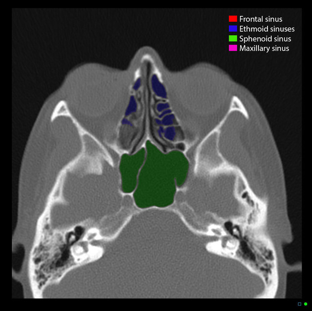
This web page presents the anatomical structures found on paranasal sinuses ct. Paranasal sinuses ct anatomy. Others are much smaller.
Your cheekbones hold your maxillary sinuses the. Welcome to interactive ct sinus anatomy. Sinuses ct brain bone windows the sphenoid sinus and ethmoid air cells are continuous with the nasal airways the mastoid air cells are continuous with the middle ear frontal sinuses ct brain bone windows.
Information about the sinus anatomy of individual patients is essential prior to a fess procedure functional endoscopic sinus surgery. The paranasal sinuses usually consist of four paired air filled spaces.

 Paranasal Sinuses Annotated Ct Radiology Case
 Startradiology
 Ct Sinuses Anatomy Quiz Radiology Case Radiopaedia Org
 Sinusitis The Cystic Fibrosis Center At Stanford
 Nasal Cavity Anatomy Physiology And Anomalies On Ct Scan
Tin Filtered 100 Kv Ultra Low Dose Ct Of The Paranasal Sinus
 A E Coronal Axial And Sagittal Ct Images Of The Bone
 Paranasal Sinus Anatomy What The Surgeon Needs To Know
 Radiologic Imaging In The Management Of Sinusitis American

 Ct Anatomy Of Para Nasal Sinuses
 Nasal Cavity Anatomy Physiology And Anomalies On Ct Scan
 Figure 27 From Paranasal Sinus Anatomy What The Surgeon
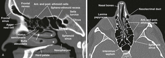
 Computed Tomography Anatomy Of The Paranasal Sinuses And
 Nose And Sinonasal Cavities Radiology Key

 Paranasal Sinus Anatomy What The Surgeon Needs To Know
 Sinusitis A Head And Neck Surgeon S Perspective
Nose And Sinonasal Cavities Radiology Key
 Startradiology
 Brain And Face Ct Interactive Anatomy Atlas
 Computed Tomography Anatomy Of The Paranasal Sinuses And

 Sinusitis In New Jersey And Philadelphia Becker Ent

 Computed Tomography Anatomy Of The Paranasal Sinuses And
 Computed Tomography Scans Of Paranasal Sinuses Before
Belum ada Komentar untuk "Ct Sinuses Anatomy"
Posting Komentar