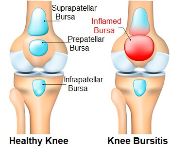
Knee Bursa Anatomy
The bursae of the knee are the fluid sacs and synovial pockets that surround and sometimes communicate with the joint cavity. Knee bursitis causes pain and can limit your mobility.
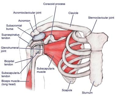
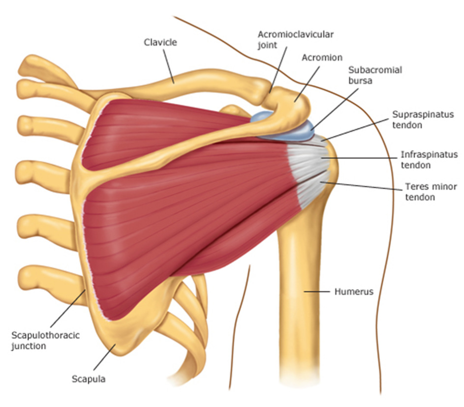 Impingement Syndrome Brisbane Knee And Shoulder
A bursa is a small fluid filled sac bounded by synovial membrane having an inner capillary layer of viscous synovial fluid.
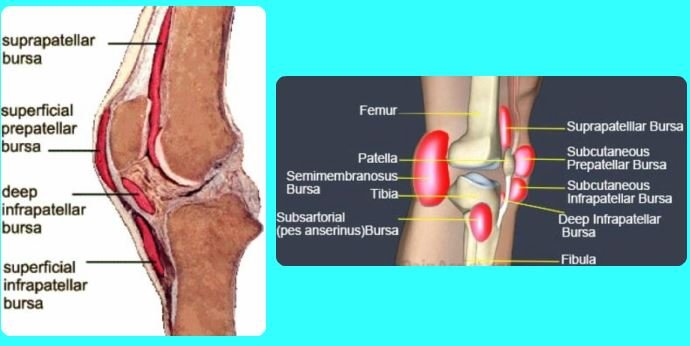
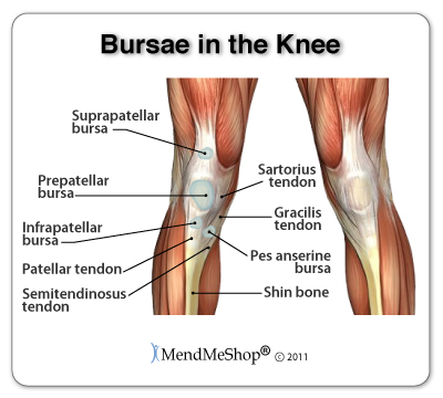
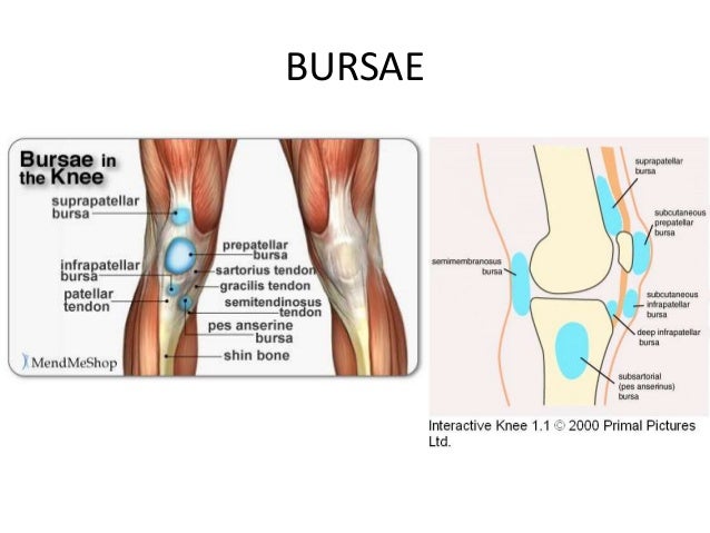
Knee bursa anatomy. Anatomy of the knee bursae a bursa is a small sac made of fibrous tissue that has an inner lining of synovial type membrane. They decrease friction and protect the fragile structures from stress. A knee bursa also known as a subcutaneous prepatellar bursa aids with movement when we walk run stretch or even cross our legs.
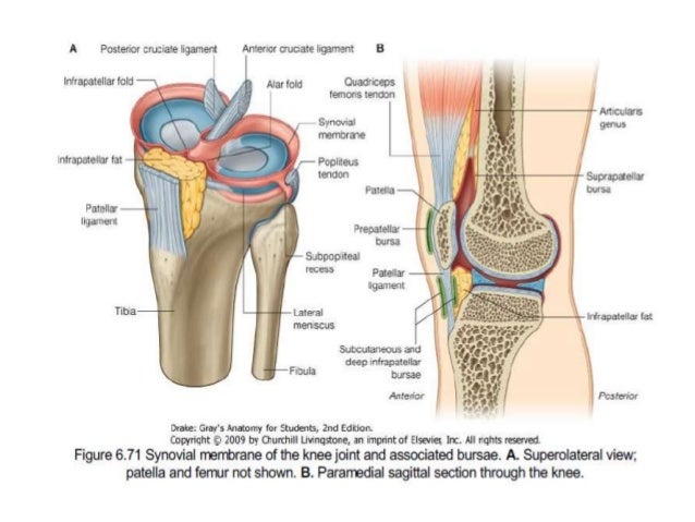
A bursa is a fluid filled structure that is present between the skin and tendon or tendon and bone. The knee contains three important groups of bursae. Between the femur and quadriceps femoris it is attached to the articularis genu muscle and communicates with the synovial cavity.
Location of anserine pes anserinus bursa on medial knee. There are four bursae anterior to the knee joint. Their function is to reduce friction caused by muscles and tendons moving against skin and bones as well as to facilitate movement.
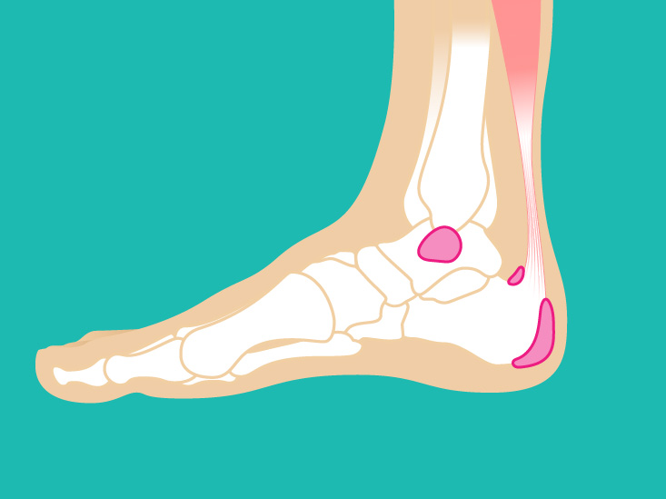
They are located over the joints and bony protuberances and may or may not interact with the joint. The main function of a bursa is to reduce friction between adjacent moving structures. The superficial one is located between the skin and the tendon and the deep one is located between the calcaneus and the tendon.
The prepatellar bursae lie in front of the patella. In the ankle two bursae are found at the level of insertion of the achilles tendon. These are located where muscles and tendons move over bony joint areas.
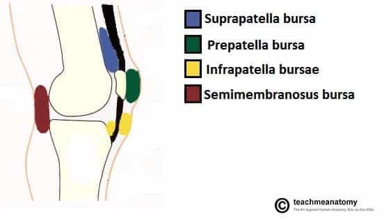
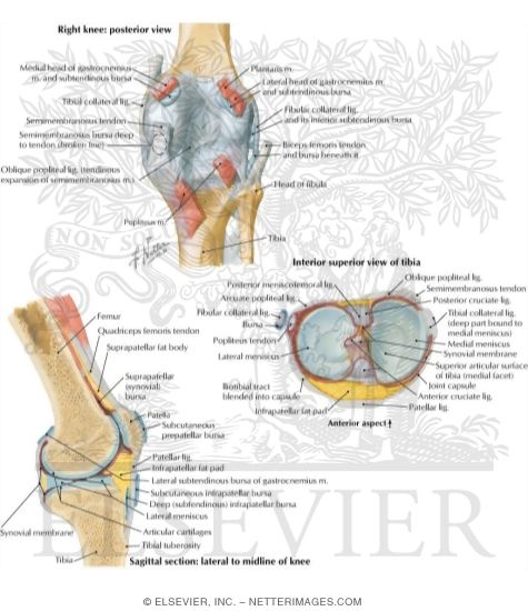
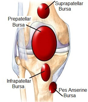
Sagittal section of right knee joint thus showing only frontal bursae. So lets have a look at knee bursitis anatomy particularly focusing on the 5 main knee bursa which are the ones that are most commonly injured. Thin walled and filled with synovial fluid they represent the weak point of the joint but also produce enlargements to the joint space.
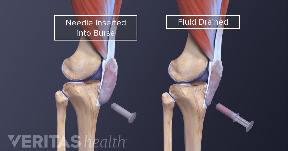
Knee bursitis is inflammation of a small fluid filled sac bursa situated near your knee joint. Bursae one is a bursa are fluid filled sacs that help cushion the knee. Between the skin and patella.
It is filled with synovial fluid or lubricant made by the membrane. Typically bursae are located around large joints such as the shoulder knee hip and elbow1 inflammation of this fluid filled structure is called bursitis. This is usually when there is excessive friction over the bursa causing it to either become inflamed or when it dries out so it no longer works properly.
 Knee Bursitis Symptoms Diagnosis Treatment
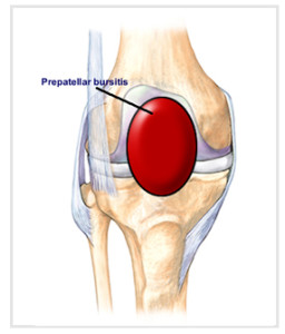
 Knee Prepatellar Bursitis
 Pin On Health Fitness

 Pin On Staying Healthy

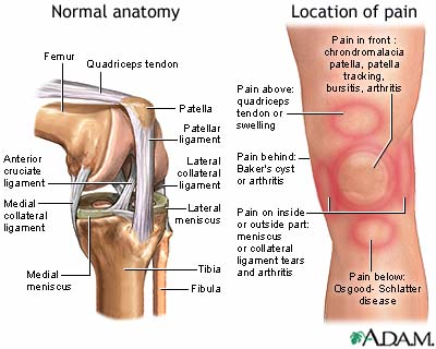 Knee Pain Medlineplus Medical Encyclopedia
 Knee Bursae Anatomy Diagram Quizlet
 Prepatellar Bursitis Treatment

 Bursitis Ankle Bursa Care And Prevention

 Dr Anurag Applied Anatomy Of Knee
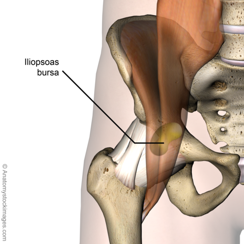 Iliopsoas Bursitis Physiopedia
Elbow Olecranon Bursitis Orthoinfo Aaos
Knee Calf Orthopedic Specialist Of Northern California
 Summit Medical Group
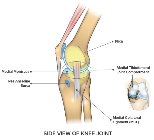 Knee Pain On The Inside Of Your Joint Causes Solutions
Trochanteric Bursitis Hip Bursitis Cleveland Clinic
 Bursitis Practice Essentials Anatomy Pathophysiology
 Prepatellar Bursitis Natural Treatment Osmo Patch Us
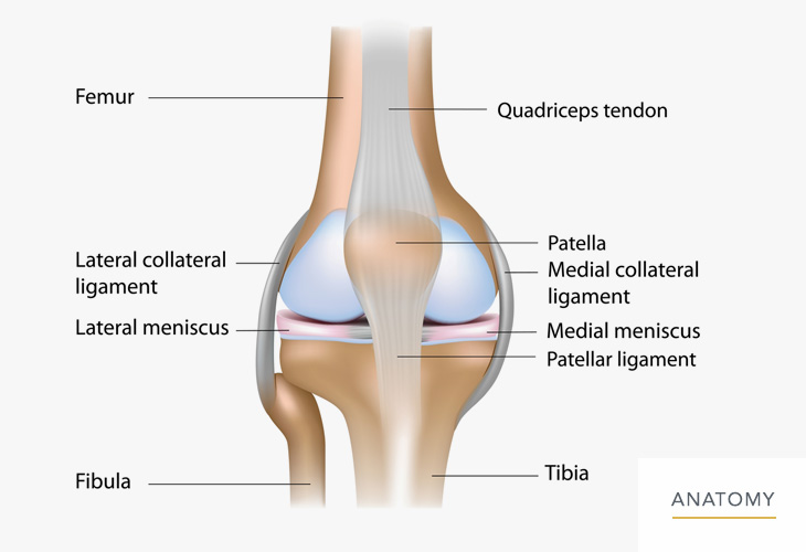
 Knee Joint Anatomy
 The Knee Joint Articulations Movements Injuries
 Knee Joint Ligaments And Bursae
 Knee Bursa Anatomy Function Injuries Knee Pain Explained
 Knee Bursae Locations Anatomy Study Com
 Knee Leg Atlas Of Anatomy
Belum ada Komentar untuk "Knee Bursa Anatomy"
Posting Komentar