Left Atrial Appendage Anatomy
Left atrial appendage function. The left atrial appendage laa is a derivative of the atrial primordium that has anatomical and physiologic variations from the left atrium la which is an extension of the embryological pulmonary vein pv bud.

Background transesophageal echocardiography tee is the diagnostic modality of choice for visualizing the left atrial appendage laa.

Left atrial appendage anatomy. A major endocrine organ it mainly produces anp atrial natriuretic peptide supply inside the heart. It is proximal to the unfastened wall of the cardiac chamber known as the left ventricle. Left atrial appendage anatomy and function.
This study defined the morphology of the laa in normal autopsy specimen hearts and considered the implications of these findings for tee studies. It acts as a reservoir for the left atrium of the heart. Short term response to sustained atrial fibrillation m weigner s katz p douglas and w manning department of medicine cardiovascular division and the harvard thorndike laboratory of beth israel deaconess medical center 330 brookline avenue boston ma 02215 usa.
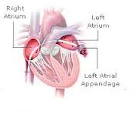
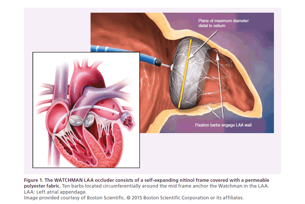
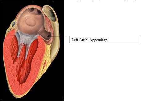
Percutaneous left atrial appendage laa closure represents a complementary option and effective treatment for patients at risk of thromboembolism especially in patients for whom it may be difficult to achieve satisfactory anticoagulation control or where anticoagulation treatment is not possible or desirable. It is a finger like projection from the main body of the la. The junction is fairly well defined by a narrowing at the orifice of the appendage.
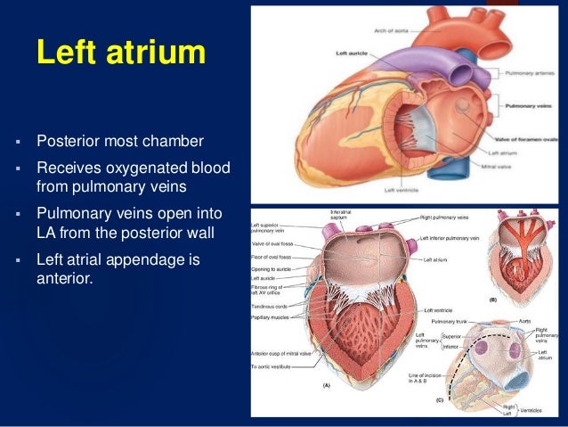
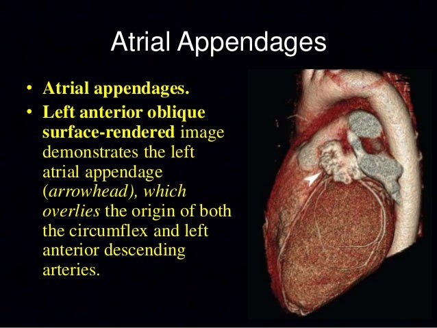
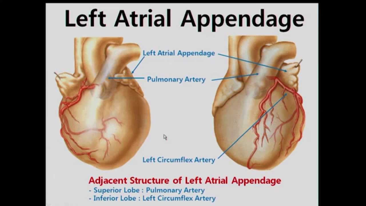
Laa appears as a windsock in appearance. The left atrial appendage laa is a pouch like projection from the main body of the left atrium lies in the atrioventricular sulcus in close proximity to the left circumflex artery the left phrenic nerve and the left pulmonary veins. The laa derives from the primordial left atrium la which is formed mainly by the adsorption of the primordial pulmonary veins and their branches 6.
Left atrial appendage anatomy.

 Left Atrial Appendage Anatomy And Imaging Landmarks
 Figure 1 From Techniques To Improve Left Atrial Appendage
 Imaging Support From 3d Tee In Left Atrial Appendage
 Watchman Left Atrial Appendage Occlusion
 Watchman Left Atrial Appendage Closure Device Treatment Animation
 Frank Silvestry Md On Twitter Would Love If You Join
 Fda Clears Watchman Device As An Alternative To
 Anatomy Of The Left Atrial Appendage Semantic Scholar
 Left Atrial Appendage Anatomy And Imaging Landmarks
Left Atrial Appendage
 The Left Atrial Appendage Anatomy Function And
 Percutaneous Left Atrial Appendage Closure A Review Of The


 Left Atrial Appendage Anatomy And Imaging Landmarks
 Outcomes Left Atrial Appendage Exclusion In Patients With
 Left Atrial Anatomy Revisited Circulation Arrhythmia And
 Anatomy And Physiology Of The Heart
 The Left Atrial Appendage In The Heart Is A Region
 Mdct Anatomy Of Heart Dr Muhammad Bin Zulfiqar
 Left Atrial Appendage Anatomy An Anatomical Imaging Review
 Left Atrial Appendage Anatomy And Imaging Landmarks
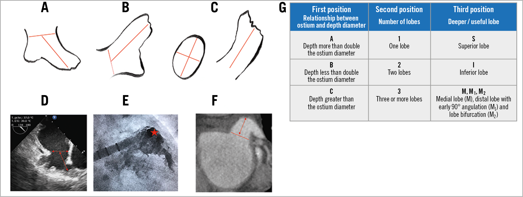 A New Practical Anatomical Classification For Left Atrial
 Left Atrial Appendage Laa Regional Anatomy And
 An Anatomical Review Of The Left Atrium Sciencedirect
 Cardiac Anatomy And Electric Mapping For Ablation Of Atrial Fibrillation
 The Amplatzer Amulet Facing The Challenge Of Left Atrial
Belum ada Komentar untuk "Left Atrial Appendage Anatomy"
Posting Komentar