Neck Of Femur Anatomy
A fractured neck of femur is classified as either intracapsular or extracapsular. The classical clinical finding is that of an externally rotated shortened leg.
 Femur Anatomy Britannica
In humans the neck of the femur connects the shaft and head at a femur upper bone of the leg or hind leg.

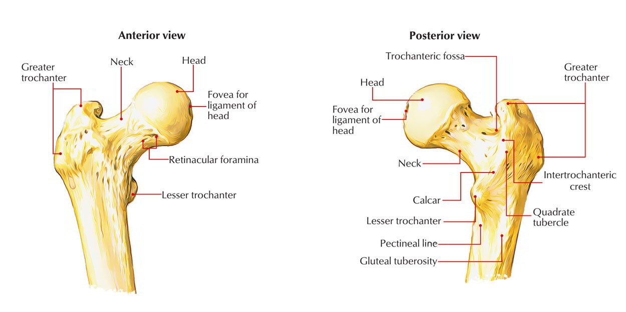
Neck of femur anatomy. In the female in consequence of the increased width of the pelvis the neck of the femur forms more nearly a right angle with the body than it does in the male. The femoral neck is strengthened by a thickening of bone called the calcar femorale present along its concavity. The neck is about is about 3 35 cms long and connects head with the shaft.
The head forms a ball and socket joint with the hip at the acetabulum being held in place by a ligament ligamentum teres femoris within the socket and by strong surrounding ligaments. There are also two bony ridges connecting the two trochanters. The proximal end consists of a head neck and two trochantersthe head faces superiorward medialward and slightly anteriorward the proximal area of the femur forms the hip joint with the pelvis.
In the adult the neck forms an angle of about 125 with the body but this varies in inverse proportion to the development of the pelvis and the stature. Head connects with the acetabulum of the pelvis to make the hip joint. Over 65000 hip fractures each year are recorded in the uk alone and they are becoming increasingly frequent due to an aging population.
The neck forms an angle with the shaft known as neck shaft angle and is about 125 in adults lesser in females. In the vast majority of cases a hip fracture is a fragility fracture due to a fall or minor trauma in someone with weakened osteoporotic bone. The angle facilitates movements of the hip joint.
A fracture of the femoral neck is classified as a type of hip fracture. With associated lengthy hospital stays and an estimated cost of over 7500 per fracture. The femoral aspect of the hip is made up of the femoral head with its articular cartilage and the femoral neck which connects the head to the shaft in the region of the lesser and greater.
It is often due to osteoporosis. A fractured neck of femur nof is a common orthopaedic presentation. Fractures of the neck of femur are very common injuries which mainly occur in elderly females with osteoporotic bones.
Proximal Femur Approach Lateral Approach Femoral Neck
 The Femur Human Anatomy
 Anatomy Of The Horse Osteology
 Anatomy Of The Hip Joint

 Femur And Hip Clinical Gate
 Bones Of The Lower Limbs Course Hero
 Hip Anatomy Recon Orthobullets
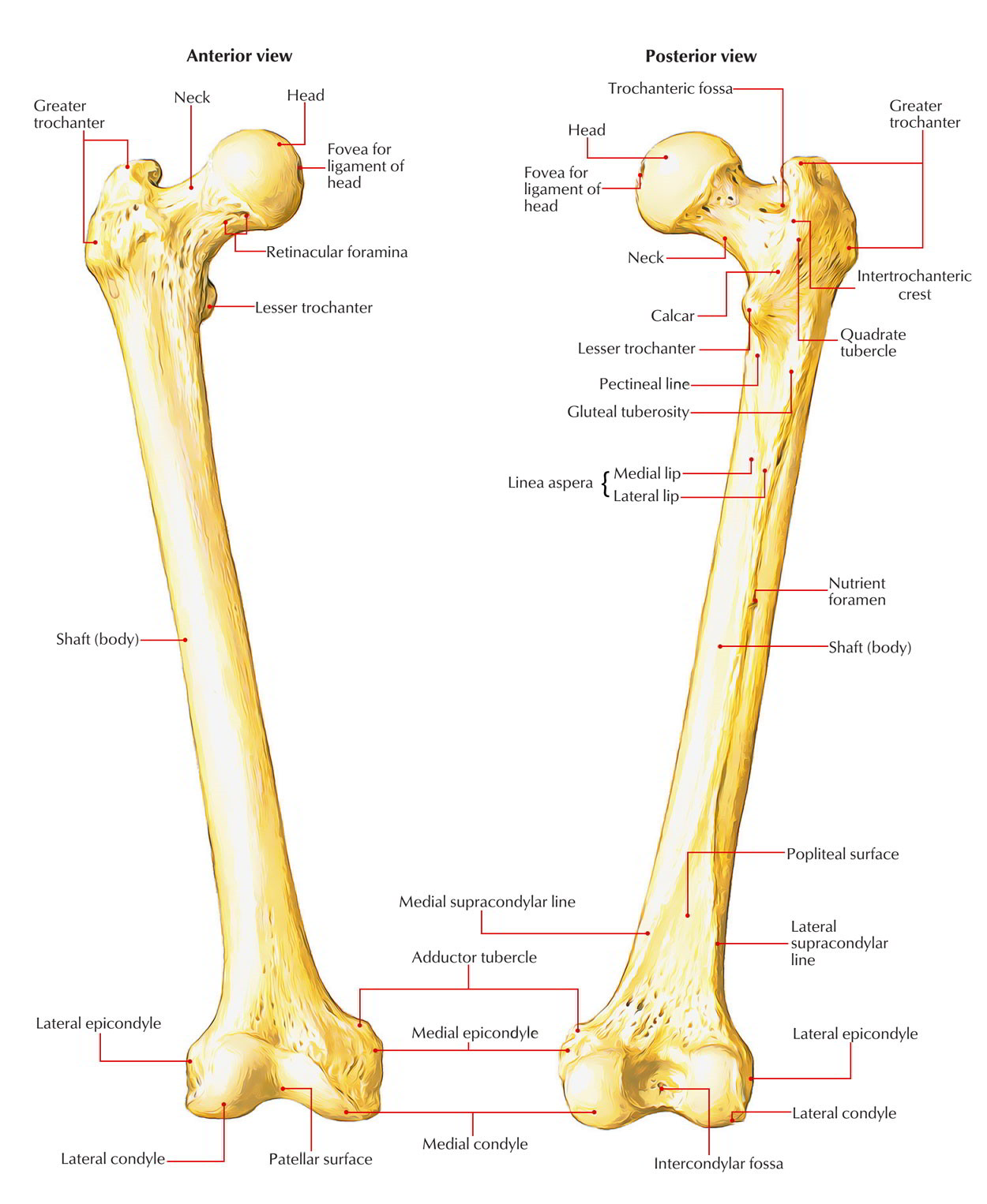
 Femur Bone Anatomy Landmarks And Muscle Attachments
 Nof Neck Of Femur Fracture Neck Fracture Greater
 Femur Wikiwand
 Femoral Neck Stress Fractures Knee Sports Orthobullets
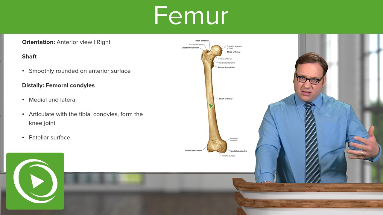 Femur Anatomy Medical Education Videos

 Bones Of The Lower Limb Anatomy And Physiology I
 The Hip Practical Office Orthopedics Accessmedicine
 Easy Notes On Femur Learn In Just 4 Minutes Earth S Lab
 Femoral Nerve Block Landmarks And Nerve Stimulator
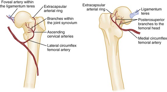
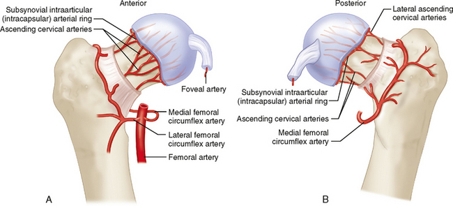
 Vascular Anatomy Of Femoral Head Neck Top Anterior
 Legg Calve Perthes Disease Vca Animal Hospital
 17 Femoral Neck Fractures Musculoskeletal Key
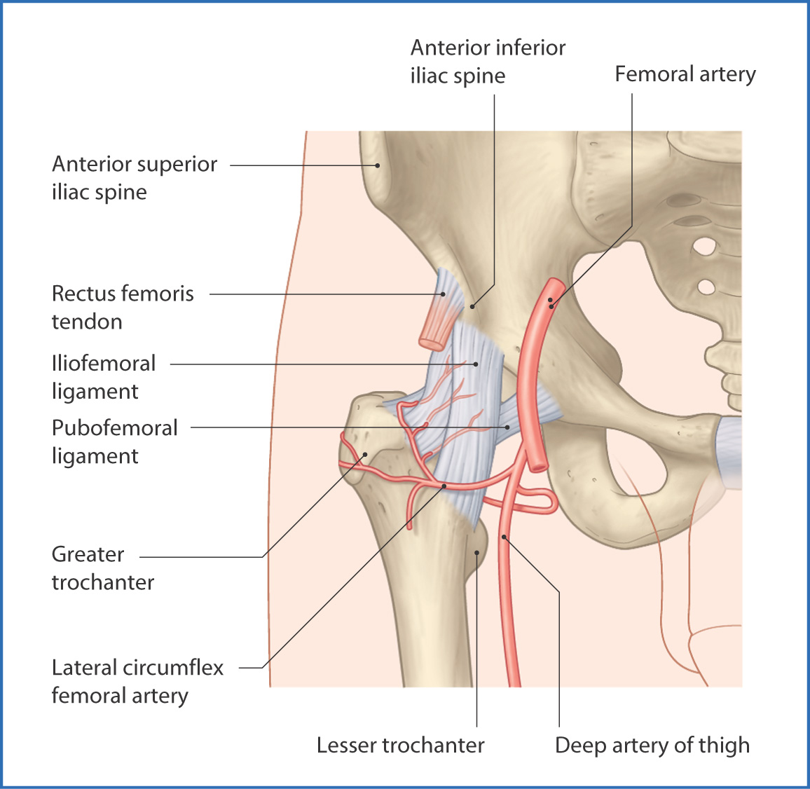
 Proximal Femur Approach Surgical Hip Dislocation Ao
![]() Femur Bone Anatomy Proximal Distal And Shaft Kenhub
Femur Bone Anatomy Proximal Distal And Shaft Kenhub
 Femur Neck Wikipedia
 Easy Notes On Femur Learn In Just 4 Minutes Earth S Lab
 Royalty Free Femur Stock Images Photos Vectors Shutterstock
Belum ada Komentar untuk "Neck Of Femur Anatomy"
Posting Komentar