Vascular Anatomy Lower Extremity
The femoral pulse can be palpated as it enters the femoral triangle midway between the anterior superior iliac spine of the pelvis and the pubis symphysis the mid inguinal point. The vascular lower extremity bachelorclass covers multiple indications that may be present in your patients examination such as cardiac output dehydration and infection which can all affect the vascular system in different ways and aggravate existing conditions.
 Presentation1 Pptx Radiological Vascular Anatomy Of The
Arterial supply of the lower limb.
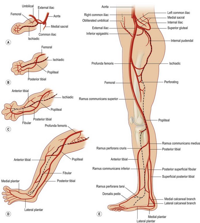
Vascular anatomy lower extremity. It runs as a single trunk from the inguinal ligament to the lower border of the popliteus where it divides into two branches the anterior and posterior tibial. Anatomy of the femur patella tibia fibula lateral and medial condyle intercondylar tubercle. Pulse points in the lower limb there are four main pulse points in the lower limb.
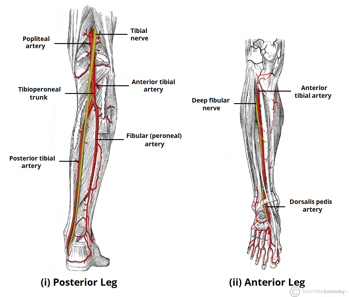
The tibioperoneal trunk divides into the posterior tibial and peroneal arteries. Peripheral vascular imaging of the lower limb. This 3d anatomy tutorial provides a basic overview on the arterial supply to the lower limb.
Anteroposterior radiology x ray of the knee. The artery which supplies the greater part of the lower extremity is the direct continuation of the external iliac. It continues to the dorsum of the foot as the dorsalis pedis artery.
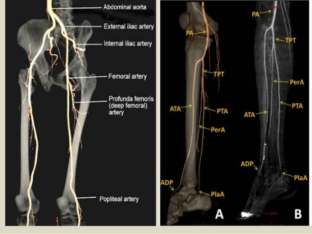
Lower extremity anatomy for blocks regionalaps rotations slides by randall j. It divides into medial and lateral plantar arteries. We preferred to use an angioct of the lower limb rather than a digital arteriography because it allowed the user to make a correlation between angiographic views of arteries with three dimensional structures.
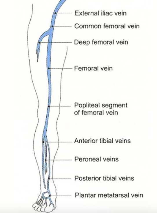
You will learn about the following structures. And the perforating veins that penetrate the muscular fascia and connect the superficial and deep veins. Femoral popliteal posterior tibial and dorsalis pedis.
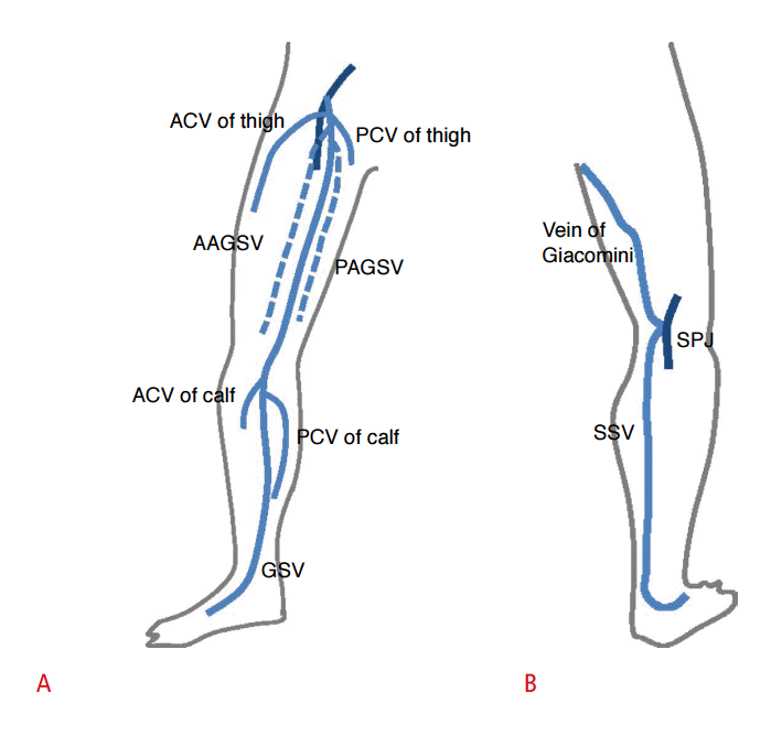
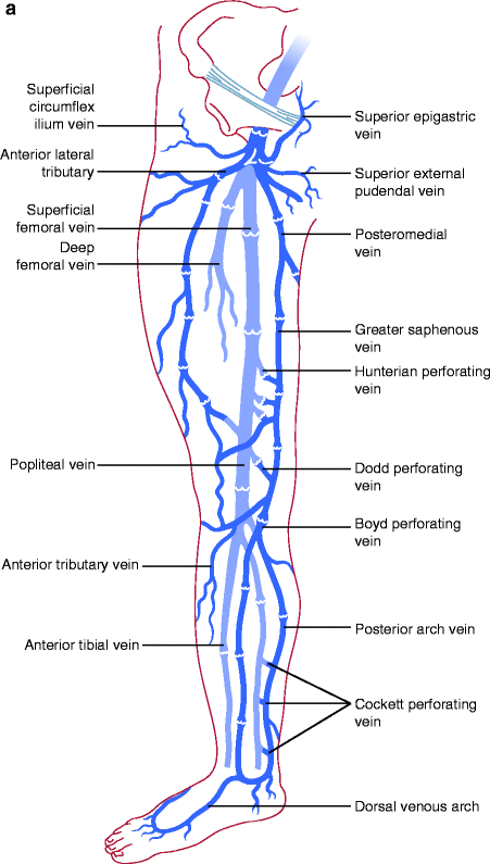
The venous system of the lower extremities includes the deep veins which lie beneath the muscular fascia and drain the lower extremity muscles. Teachme anatomy part of the teachme series the medical information on this site is provided as an information resource only and is not to be used or relied on for any diagnostic or treatment purposes. External iliac artery.
The superficial veins which are above the deep fascia and drain the cutaneous microcirculation. The posterior tibial artery passes downwards and behind the medial malleolus.
 Arteries Of The Lower Limb Thigh Leg Foot Teachmeanatomy
 Vascular Surgery Lower Extremity Artery Diagram Wiring Diagram
 Module 2 Lower Extremity Orthopedic Imaging
 Ultrasonography
 Arterial Anatomy Of The Extremities Radiology Key
 Surgical Bypass Society For Vascular Surgery
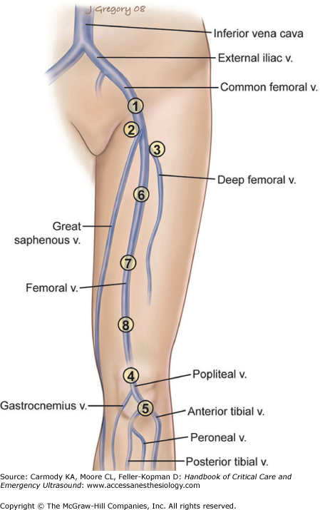 Chapter 15 Ultrasound For Deep Venous Thrombosis Handbook
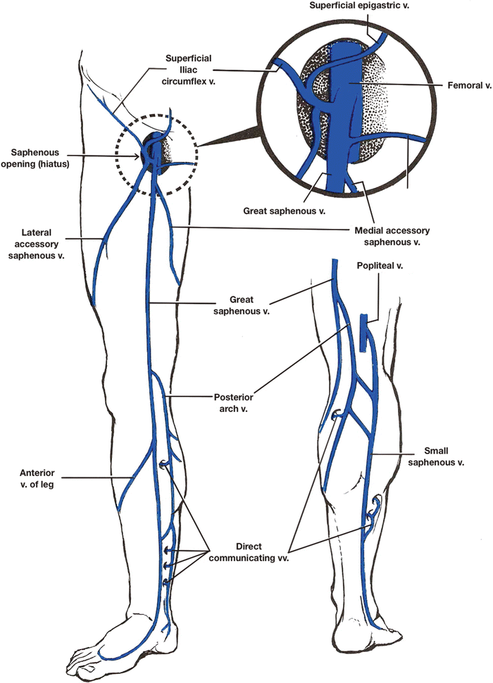
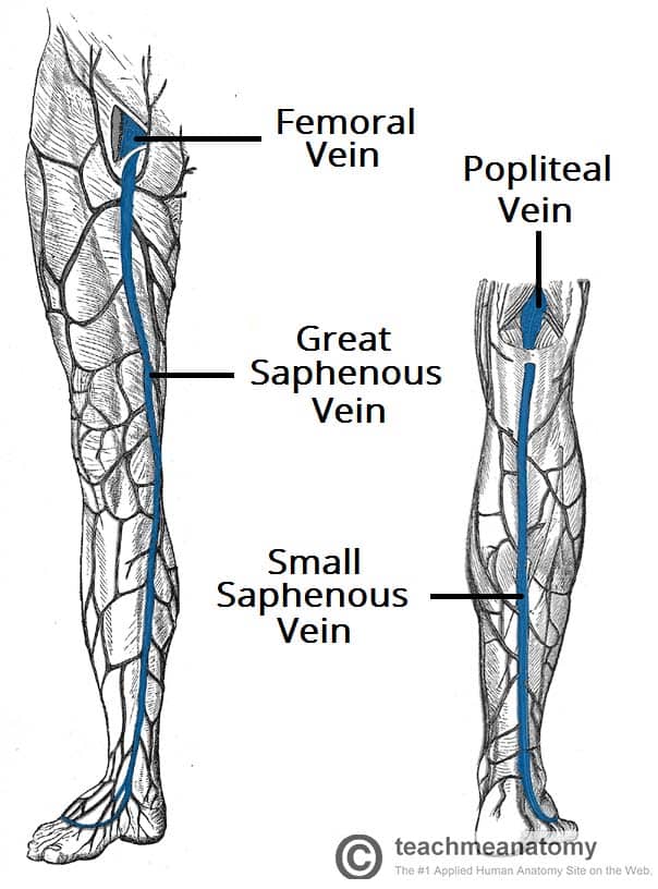 Venous Drainage Of The Lower Limb Teachmeanatomy
 Circulatory System Of The Lower Extremities Medical Addicts
![]() Lower Extremities Arteries And Nerves Anatomy Branches
Lower Extremities Arteries And Nerves Anatomy Branches
 Vascular Anatomy Of The Left Lower Extremity With Occlusion
![]() Lower Extremities Arteries And Nerves Anatomy Branches
Lower Extremities Arteries And Nerves Anatomy Branches

 Vascular Anatomy Of The Lower Limbs Springerlink
Comprehensive Lower Extremity Anatomy Plastic Surgery Key
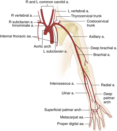
 The Arteries Of The Lower Extremity Human Anatomy
 Ultrasound Guided Saphenous Adductor Canal Block Nysora
 Blood Supply To The Foot Foot Ankle Orthobullets

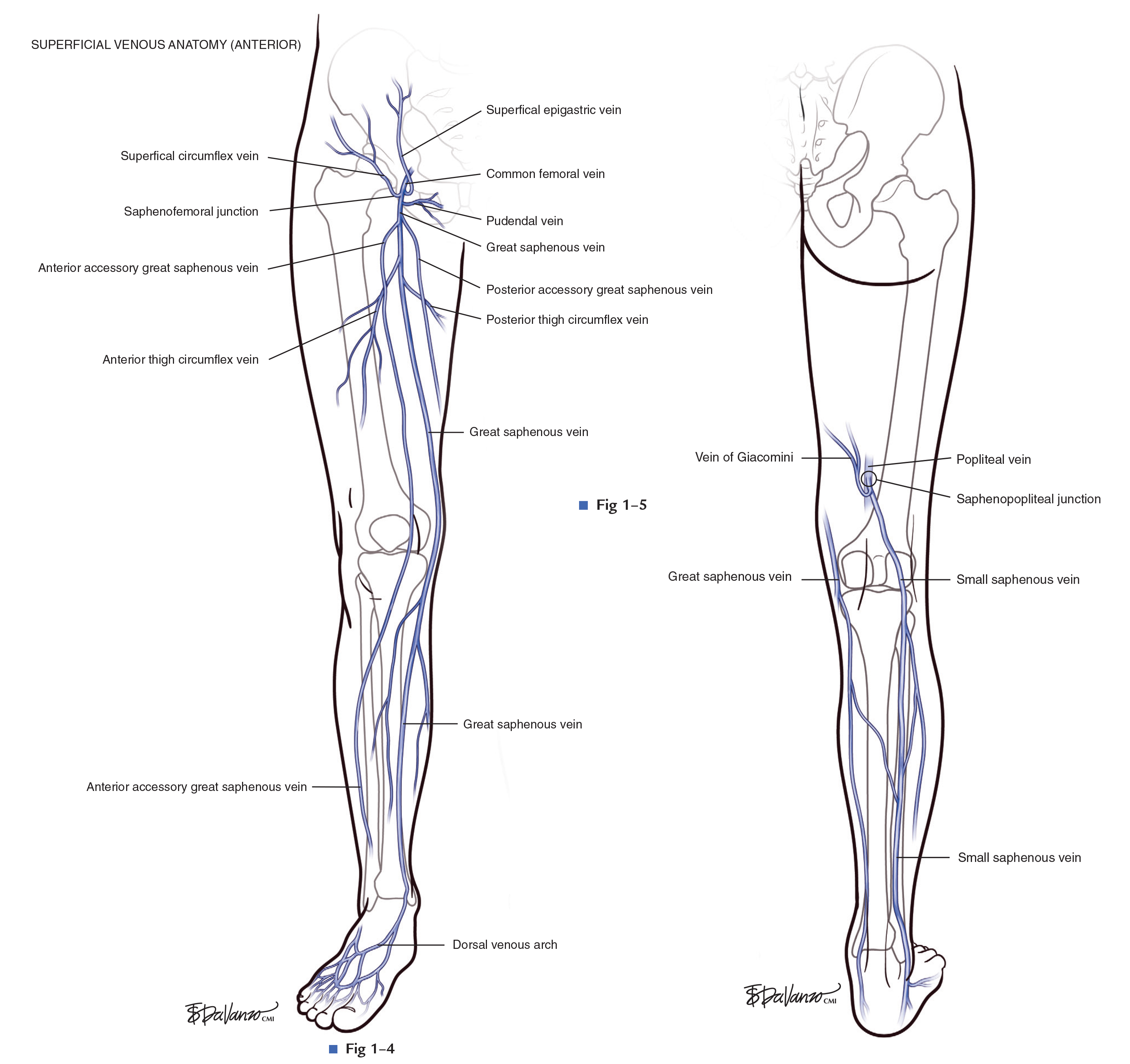
 Anatomy Of The Lower Extremity Veins Varicose Veins

 Lower Extremity Venous Ablation And Sclerotherapy Springerlink
 Deep Venous Thrombosis Dvt Core Em
Belum ada Komentar untuk "Vascular Anatomy Lower Extremity"
Posting Komentar