Anatomy Of Fetus
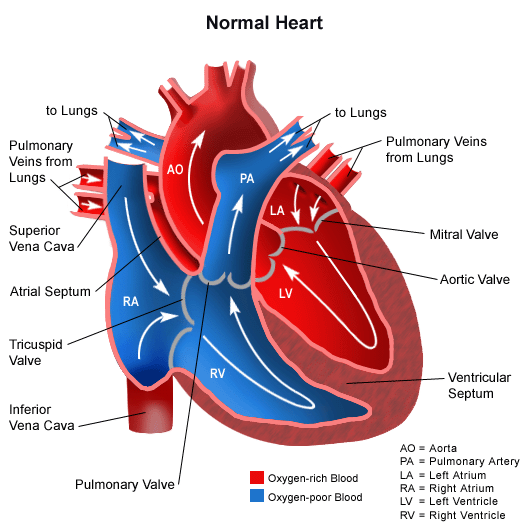
A thin walled sac that surrounds the fetus during pregnancy. In this case the blood flowing to the fetal heart is oxygenated because it comes from the placenta.
 Fetus In Utero Anatomy Watercolor Splash
When a level 2 ultrasound is done.

Anatomy of fetus. Most anatomy scans are performed in the second trimester of pregnancy typically at 20 weeks but they can be done anytime between 18 weeks and 22 weeks. An organ shaped like a flat cake. The anatomy scan is a level 2 ultrasound which is typically performed on pregnant women between 18 and 22 weeks.
Brain ventricles choroid plexus mid brain posterior fossa cerebellum cisterna magna. However a fetus is characterized by the presence of all the major body organs though they will not yet be f. Uterus also called the womb the uterus is a hollow pear shaped organ located in a womans lower abdomen between the bladder and the rectum that sheds its lining each month during menstruation and in which a fertilized egg ovum becomes implanted and the fetus develops.
Skull shape integrity bpd and hc measurements. A fetus or foetus is the unborn offspring of an animal that develops from an embryo. Fetus in utero amniotic sac.
Those who want to can find out the sex of the baby if desired. Remember that veins carry blood toward the heart. Heart rate rhythm 4 chamber views.
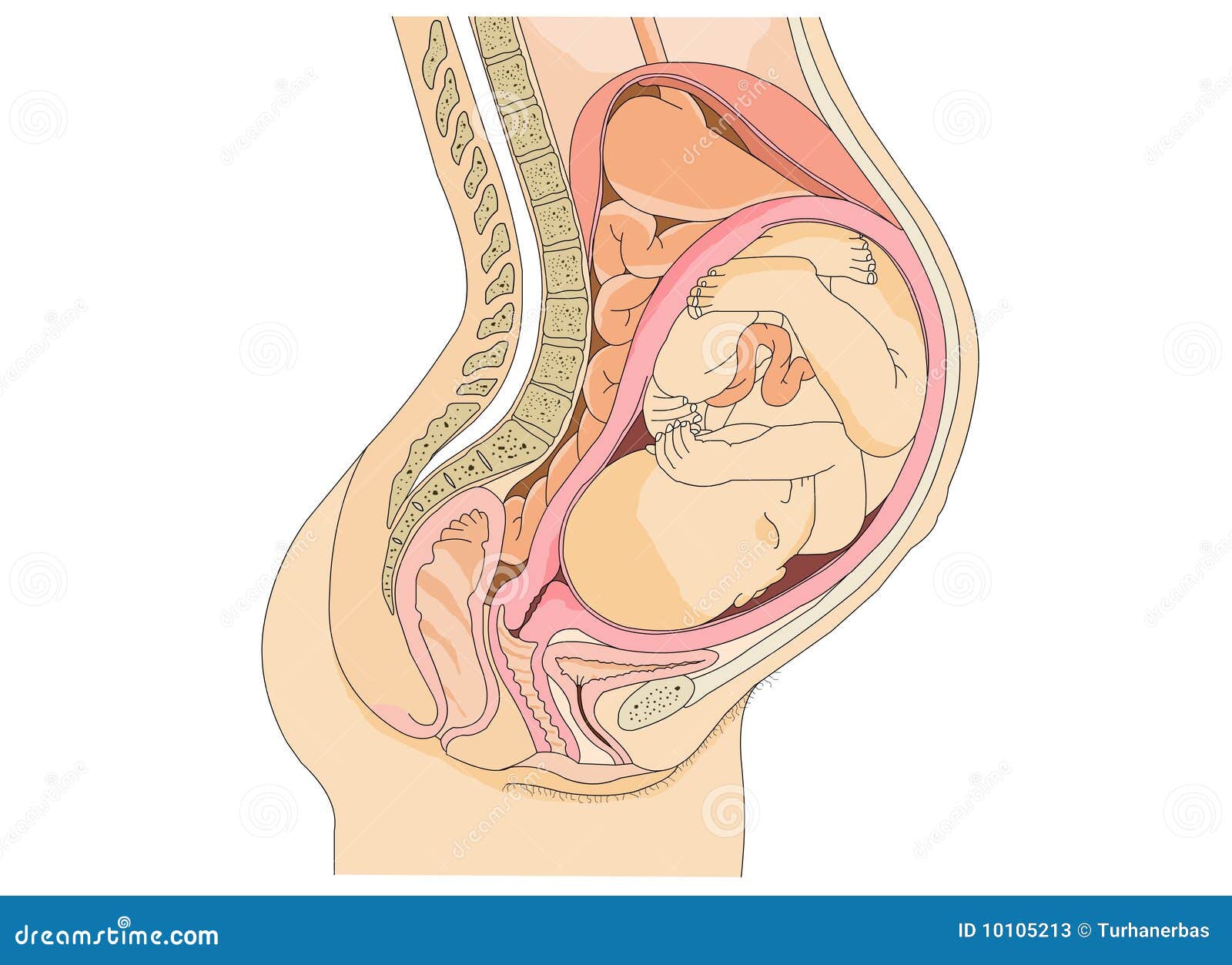
Fetus in utero amniotic sac. The following fetal parts are checked during the anatomy ultrasound. The placenta provides the fetus with necessary oxygen and nutrients via the umbilical vein.
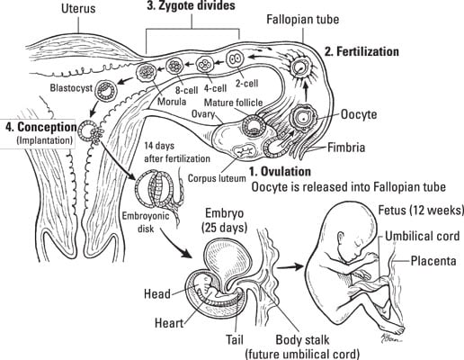
Prenatal development is a continuum with no clear defining feature distinguishing an embryo from a fetus. Systematic study of normal fetal cardiac anatomy arrangement axis and position the first step of the fetal cardiac interrogation is to describe the arrangement and location of the heart in relation to the overall arrangement of the fetus. It only grows.
Neck nuchal fold thickness. The respiratory system is immature and cannot yet oxygenate blood on its own. In human prenatal development fetal development begins from the ninth week after fertilisation and continues until birth.
The lower part of the uterus that extends into the vagina. The fetal period lasts from the ninth week of development until birth. Following embryonic development the fetal stage of development takes place.
The fetal circulatory system becomes much more specialized and efficient than its embryonic counterpart. An unborn baby from the 8th week after fertilization until birth. During this period male and female gonads differentiate.
Vagina the part of the female genitals. If you have a condition that needs to be monitored such as carrying multiples you may have more than one detailed ultrasound.
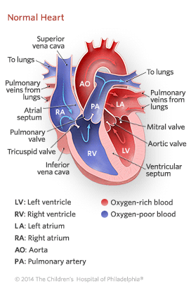 Blood Circulation In The Fetus And Newborn Children S
 Human Development Before Birth Dummies
 Fetus Baby In Womb Anatomy Stock Illustration Illustration
 Pregnant Anatomy With Fetus Clip Art K17266891 Fotosearch
 Altay Scientific Anatomy Model Pregnancy Pelvis With Mature
The Fetal Period Boundless Anatomy And Physiology
 Pin On Female
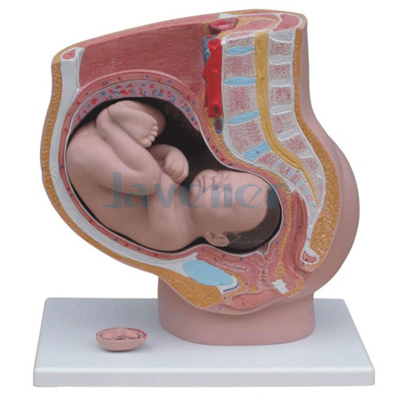 Details About Human Female Pelvic Section Pregnancy Anatomical Model Anatomy Fetus 40 Weeks
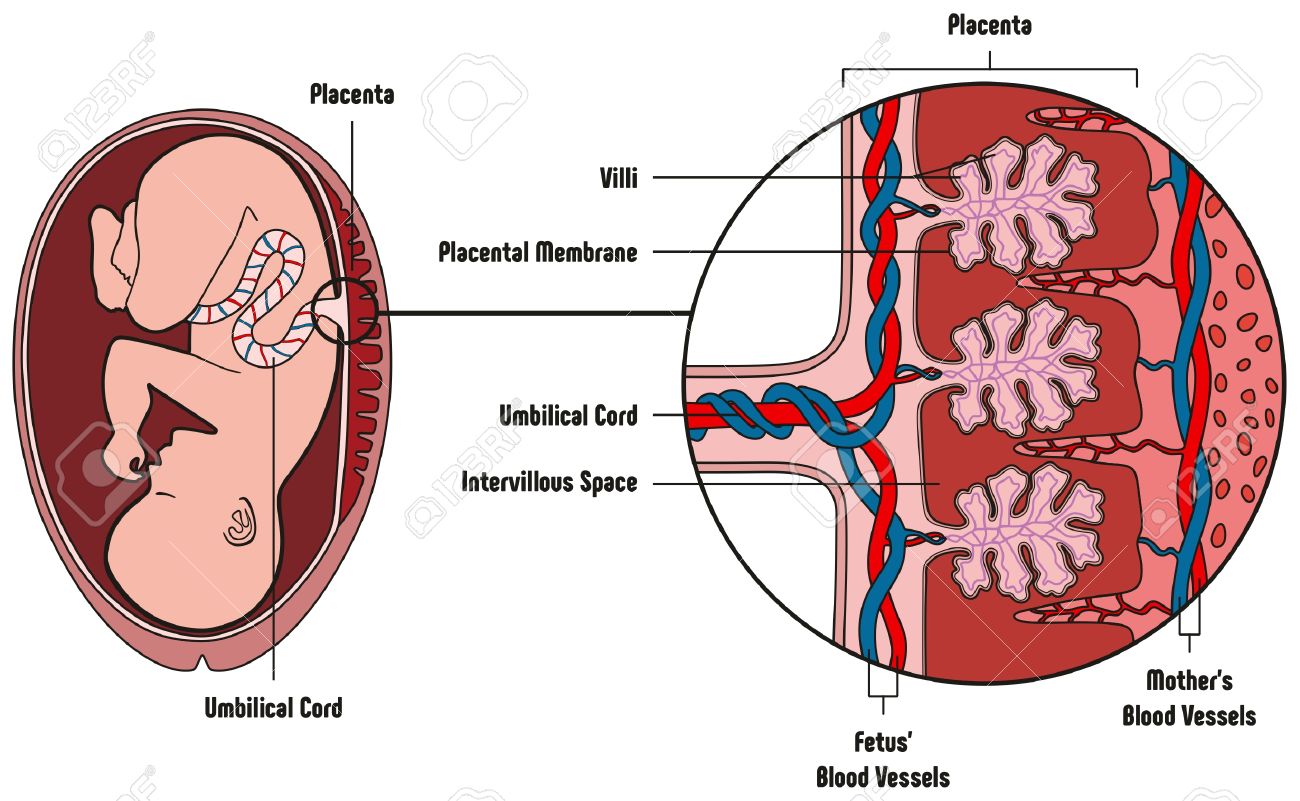 Human Fetus Placenta Anatomy Diagram With All Part Including
 Anatomy Chapter One Pregnancy Understanding Birth
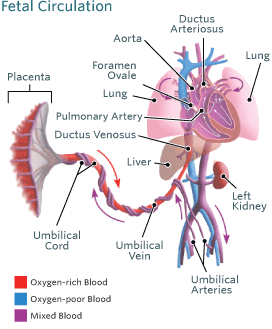
 Fetal Circulation Etc Remake Anatomy Physiology Bio 212
 Uterus Growth During Pregnancy
 Pregnant Woman Anatomy And Fetus Isolaed On White
 Pregnant Woman Anatomy Fetus Isolaed On Stock Photo Edit
 Pin On Natural Birthing

 Anatomy Pregnancy And Birth Models
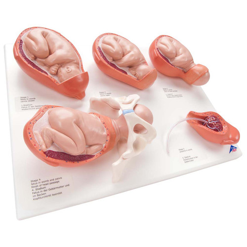
 Pregnancy Models Series 5 Embryo Fetus Models On A Base 3b Smart Anatomy
 Fetus Baby In Womb Anatomy Stock Illustration Illustration
 Pregnancy Pregnant Vector Photo Free Trial Bigstock
 11205 01x Fetal Membranes Anatomy Exhibits
 Amazon Com Axis Scientific Anatomy Model Of Pregnancy
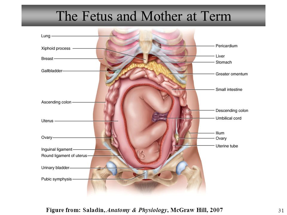
 Anatomy Of Full Term Pregnancy
:max_bytes(150000):strip_icc()/GettyImages-90113438-56cd30dc3df78cfb37a30eb2.jpg) The Role Of The Mucus Plug In Pregnancy And Labor
The Role Of The Mucus Plug In Pregnancy And Labor

 Pregnant Anatomy And The Fetus Stock Vector Illustration
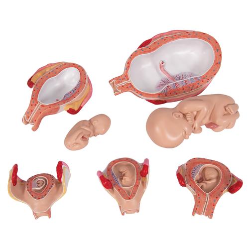
 Anatomical Human Fetal Development Model Baby Fetus Foetus Pregnancy Anatomy Science Toy
 Anatomy And Physiology Pregnancy Growth And Development
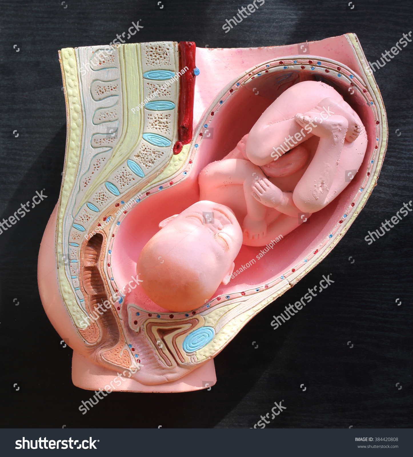 Pregnant Woman Anatomy Fetus Stock Photo Edit Now 384420808
 Blood Circulation In The Fetus And Newborn Children S
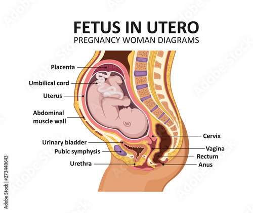 Fetus In Utero Pregnancy Women Diagrams Pregnant Female
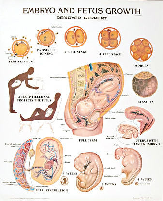 Embryo And Fetus Growth Anatomy Poster
 Details About 3b Scientific Xl004 Spare Fetus For L10 6 Anatomical Model Anatomy
Belum ada Komentar untuk "Anatomy Of Fetus"
Posting Komentar