Anatomy Of A Molar Tooth
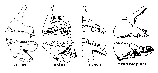
Most people start off adulthood with 32 teeth not including the wisdom teeth. Teeth between the canines and molars.
 Human Tooth Anatomy Vector Diagram Of Healthy Molar
The tissue composition of a tooth is only found within the oral cavity and is limited to the dental structures.
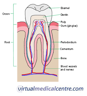
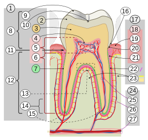
Anatomy of a molar tooth. Carabelli documented the particular anatomy of maxillary molars as early as 1844. The pulp dentin and enamel. Pulp this is the soft tissue found in the center of all.
Each tooth has several distinct parts. The development appearance and classification of teeth fall within its purview. The third rearmost molar in each group is called a wisdom toothit is the last tooth to appear breaking through the front of the gum at about the age of 20 although this varies from individual to individual.
Premolars 8 total. The root is the part of the tooth that extends into the bone and holds. Tooth anatomy types of teeth.
Each tooth is an organ consisting of three layers. Twelve molars are usually present in an adult human. Here is an overview of each part.
Explore the interactive 3 d diagram below to learn more about teeth. Wisdom teeth or third molars 4 total. The anatomy of maxillary molars is very complex and the root canal treatment of this particular group of teeth represents a major challenge for dentists.
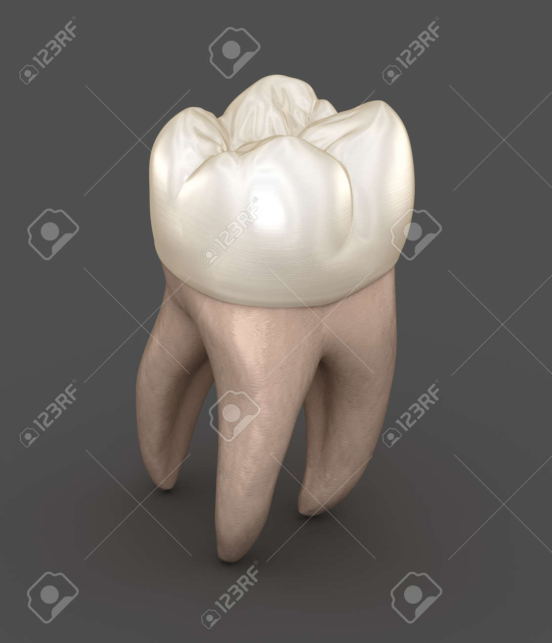
The mandible and maxilla like most bones in the human body have a core of less dense cancellous bone wrapped in an outer layer of more dense alveolar bone. Enamel this is the outer and hardest part of the tooth that has the most mineralized tissue in. A molar tooth is located in the posterior back section of the mouth.
Tiny blood vessels and nerve fibers enter the pulp through small holes in the tip of the roots to support the hard outer structures. Anatomy of the tooth the tooth is one of the most individual and complex anatomical as well as histological structures in the body. It is found in most mammals that use their posterior teeth to grind food.
These teeth erupt at around age 18 but are often surgically removed to prevent displacement of other teeth. Anatomy and physiology of the teeth in the mouth the bone holding the bottom row of teeth is the mandible and the bone holding the top row of teeth is the maxilla. Flat teeth in the rear of the mouth best at grinding food.
In humans the molar teeth have either four or five cuspsadult humans have 12 molars in four groups of three at the back of the mouth. The pulp of the tooth is a vascular region of soft connective tissues in the middle of the tooth. The function of teeth as they contact one another falls elsewhere under dental occlusion tooth formation begins before birth.
Dental anatomy is a field of anatomy dedicated to the study of human tooth structures. Most often the. Numerous subsequent publications discussed the complexity of maxillary molar anatomy.
Dentin this is the layer of the tooth under the enamel. Molars 8 total.
 Steven G Starr Dds Endodontics Root Canals Saint
 Human Anatomy Molar Enlargement Model Healthy Large Tooth Structure Oral Dental Teaching Mold Decoration
 Orthodontist Human Tooth Anatomy Vector Infographics With
The Permanent Mandibular Molars Dental Anatomy Physiology
:background_color(FFFFFF):format(jpeg)/images/library/10657/Tooth_Thumbnail_v01__1_.png) Tooth Anatomy Names Types Structure Arteries Nerves
Tooth Anatomy Names Types Structure Arteries Nerves
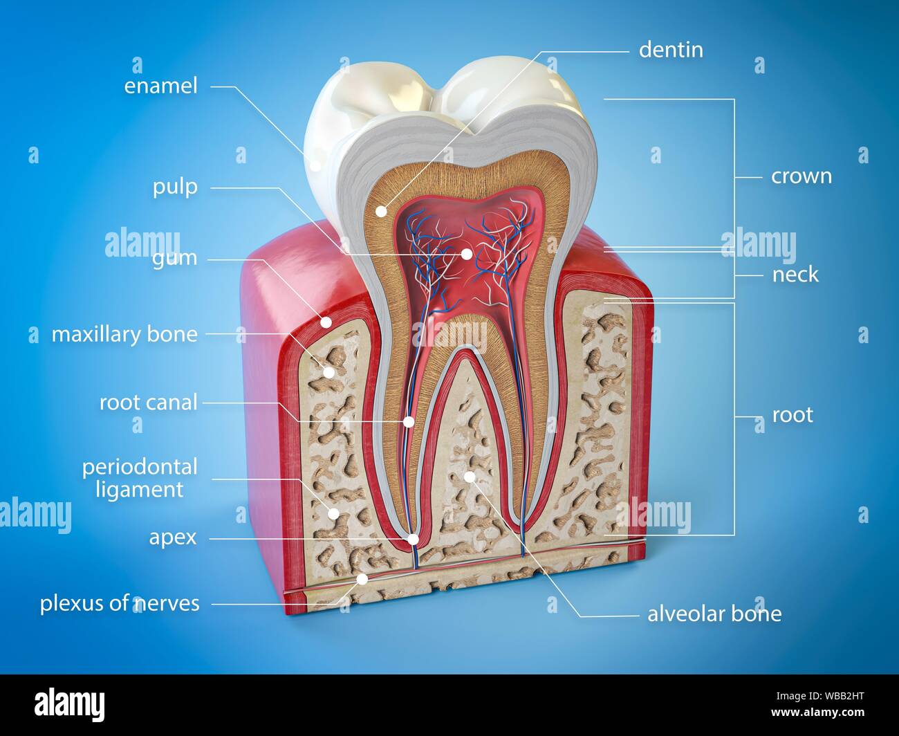 Molar Tooth Cross Section Stock Photos Molar Tooth Cross
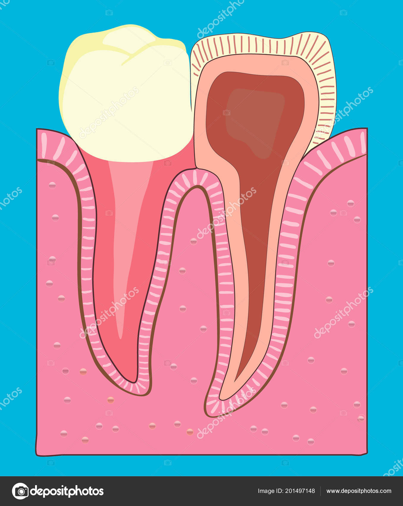 Anatomy Of The Molar Tooth Stock Vector C Ashpin Tyev List
 Primary Dentition An Overview Of Dental Anatomy
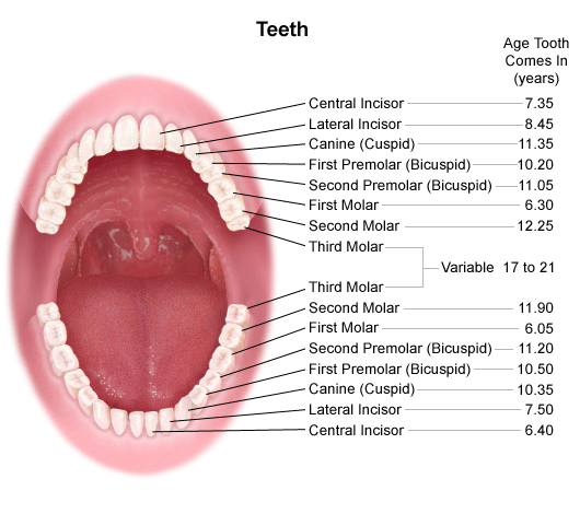 Anatomy And Development Of The Mouth And Teeth
 Tooth Anatomy Section Of A Human Molar
 Permanent Dentition An Overview Of Dental Anatomy
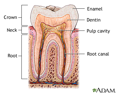 Tooth Anatomy Medlineplus Medical Encyclopedia Image
 Teeth Anatomy Adult Teeth Permanent Dentition
 Chipped Loose And Broken Teeth 101 Dear Dr Christina
 Tooth Types Patches Discover Fishes
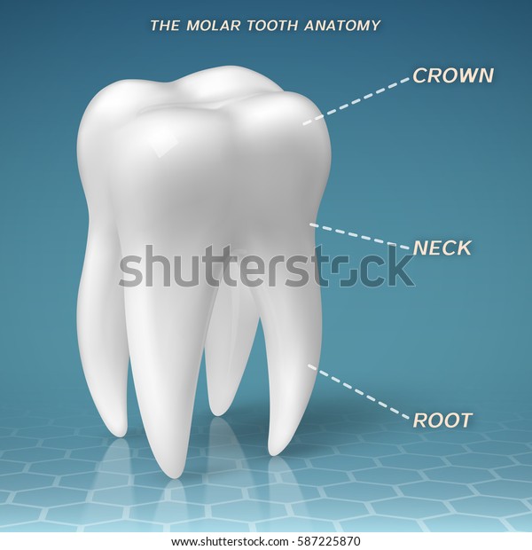 Molar Anatomy Crown Neck Root Tooth Stock Vector Royalty
Vc Dental Tooth Anatomy Education
 Molar Anatomy Shared By Dr Gregory Bowen San Antonio
 Children Teeth Anatomy Shows Eruption And Shedding Time Dental
Review Of Tooth Morphology Dental Anatomy Physiology And
/toothanatomy2.jpg)



Belum ada Komentar untuk "Anatomy Of A Molar Tooth"
Posting Komentar