Anatomy Of Hip Area
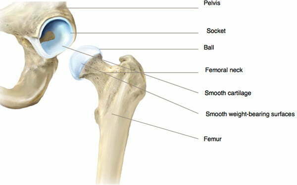
Captioned anatomical structures of the gluteal area buttocks ligaments. The hip joint is the uppermost part of the leg where the head of the thigh bone femur fits into the socket of the pelvis.
 1 Anatomy Of Hip Joint Adapted From 33 Download
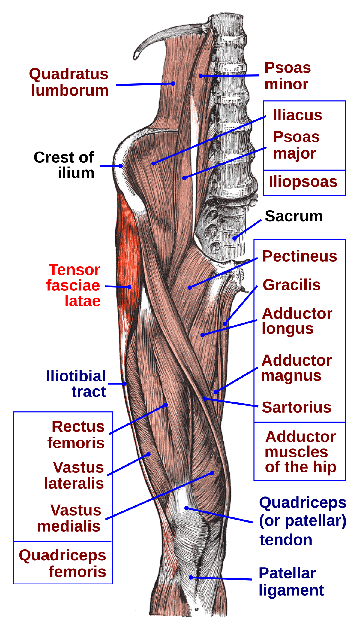
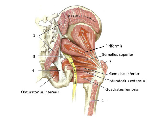
Muscle and tendon anatomy of the hip adductors gluteal muscles or buttocks hamstring muscles femoral muscle quadrices.
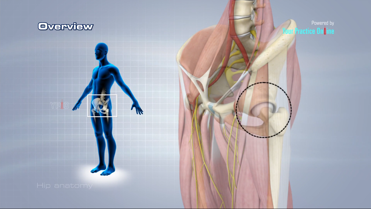
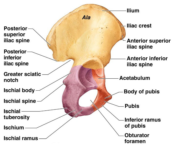
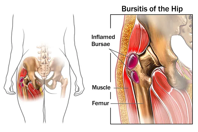
Anatomy of hip area. The hip is the bodys second largest weight bearing joint after the knee. Hip bones there are two hip bones one on the left side of the body and the other on the right. Hip pain may result from inflammation degeneration or injury to structures and tissues within the hip joint.
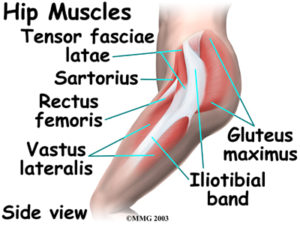
The hip joint is the articulation of the pelvis with the. Rectus femoris muscle one of the quadriceps muscles on the front of your thigh. Nerves and vessels supply the muscles and bones of the hip.
Iliopsoas muscle a hip flexor muscle that attaches to the upper thigh bone. A round cup shaped structure on the os coxa known as the acetabulum forms the socket for the hip joint. Some of the other muscles in the hip are.
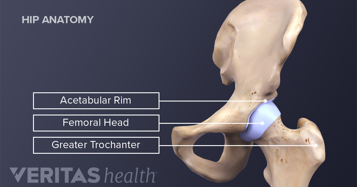
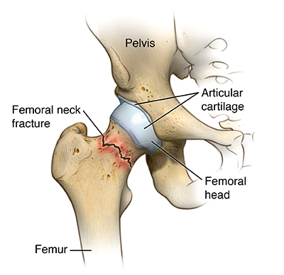
The hip is located where the head of the femur or thighbone fits into a rounded socket of the pelvis. It is a ball and socket joint at the juncture of the leg and pelvis. It is a ball and socket joint at the juncture of the leg and pelvis.
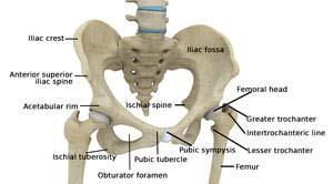
Together they form the part of the pelvis called the pelvic girdle. Hip anatomy the hips unique anatomy enables it to be both extremely strong and amazingly flexible so it can bear weight and allow for a wide range of movement. The ball is the femoral head and the socket is the acetabulum.
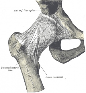
The hip region is located lateral and anterior to the gluteal region inferior to the iliac crest and overlying the greater trochanter of the femur or thigh bone. Its the need for such a high degree of stabilization of the joint that limits movement. Contains the main ligaments of the hip and its associated area iliofemoral ishciofemoral ligament and pubofemoral ligaments ligament of the head of the femur or round ligament.
Muscles play an important role in the health and well being. Anatomy of the hip. In adults three of the bones of the pelvis have fused into the hip bone or acetabulum which forms part of the hip region.
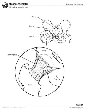
The hip joint see the image below is a ball and socket synovial joint. The hip bones join to the upper. The hip joint is a ball and socket synovial joint formed between the os coxa hip bone and the femur.
If you think of the hip joint in layers the deepest layer is bone then ligaments of the joint capsule and the tendons and muscles are on top. Adductor muscles on the inside of your thigh.
 Why People Have To Squat Differently The Movement Fix
 Hip Surgeon Oakland Ca Hip Surgery San Francisco Ca
 How To Work And Use Your Glute Muscles Correctly In Yoga
 Hip Anatomy Pictures Function Problems Treatment
 Hip Joint Anatomy Overview Gross Anatomy
 The Truth About Cracking Popping Joints Yoga Journal
:background_color(FFFFFF):format(jpeg)/images/library/11030/Hip_and_thigh_1.png) Hip And Thigh Bones Joints Muscles Kenhub
Hip And Thigh Bones Joints Muscles Kenhub
 Ligaments Tendons And Muscles Of The Hip Joint Naples
 Hip Pain Symptoms Treatment Causes Exercises Relief
 Illitibial Band Syndrome Symptoms Treatment Exercises
 Tensor Fasciae Latae Muscle Wikipedia
 Hip Joint Anatomy Hip Bones Ligaments Muscles
Hip Labral Tear Cleveland Clinic
 Functional Anatomy Of The Small Pelvic And Hip Muscles
 Hip Bone Anatomy Or Pelvic Bone Ilium Pubis Ischium Bone
 Bones Of The Pelvis Hip Bones Anatomy Tutorial
 Muscles Of The Hips And Thighs Human Anatomy And




Belum ada Komentar untuk "Anatomy Of Hip Area"
Posting Komentar