Foot Anatomy Image
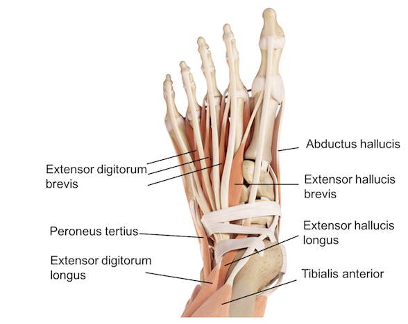
Anatomy of the foot. The foot is the lowermost point of the human leg.
 Facts About Feet Anatomy Snippets Complete Anatomy
See an illustration picture of and learn about anatomical structures in the body like the foot in the emedicinehealth image collection gallery.

Foot anatomy image. Download foot anatomy stock photos. The foots shape along with the bodys natural balance keeping systems make humans capable of not only walking but also running climbing. Learning your foot anatomy is important especially to know which bone is currently causing foot pain.
Find foot anatomy stock images in hd and millions of other royalty free stock photos illustrations and vectors in the shutterstock collection. Also for students and health professionals it is critical to understand the foot anatomy which basically improves your knowledge of bone position ligament attachment and the way tendons run on the foot bones. Feet are incredibly complex comprised of 28 bones 30 joints and more than 100 ligaments.
In humans the foot is one of the most complex structures in the body. Small alterations in the foot can affect its whole structure and cause pain when walking. The foot is an extremely complex anatomic structure made up of 26 bones and 33 joints that must work together with 19 muscles and 107 ligaments to execute highly precise movements.
The end of the leg on which a person normally stands and walks. Picture of foot anatomy detail foot. See more ideas about ankle anatomy anatomy and anatomy and physiology.
It is made up of over 100 moving parts bones muscles tendons and ligaments designed to allow the foot to balance the bodys weight on just two legs and support. Webmd image collection reviewed by carol dersarkissian on may 18 2019. Thousands of new high quality pictures added every day.
Topics a z slideshows images quizzes supplements medications. Affordable and search from millions of royalty free images photos and vectors. The foot is a part of vertebrate anatomy which serves the purpose of supporting the animals weight and allowing for locomotion on land.
Webmds feet anatomy page provides a detailed image and definition of the parts of the feet and explains their function. Jul 5 2019 explore hgbarreses board ankle anatomy on pinterest. Picture of foot anatomy detail the feet sit at the end of the legs and we use them for standing and walking.
 3b Scientific A31 1 Foot Skeleton Flexibly W Portions Of Tibia Fibula 3b Smart Anatomy
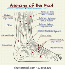 Foot Anatomy Images Stock Photos Vectors Shutterstock
 Muscles Of The Lower Leg And Foot Human Anatomy And
 Facts About Feet Anatomy Snippets Complete Anatomy
 General International Association For Dance Medicine Science
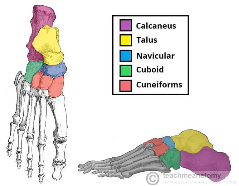 Bones Of The Foot Tarsals Metatarsals Phalanges
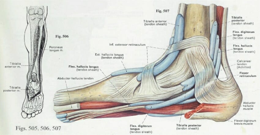 Foot Anatomy Bones Ligaments Muscles Tendons Arches
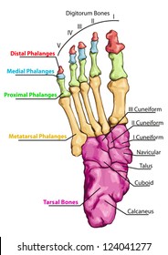 Foot Anatomy Images Stock Photos Vectors Shutterstock
 Foot Anatomy Muscular And Skeletal Anatomy Of Ankle And
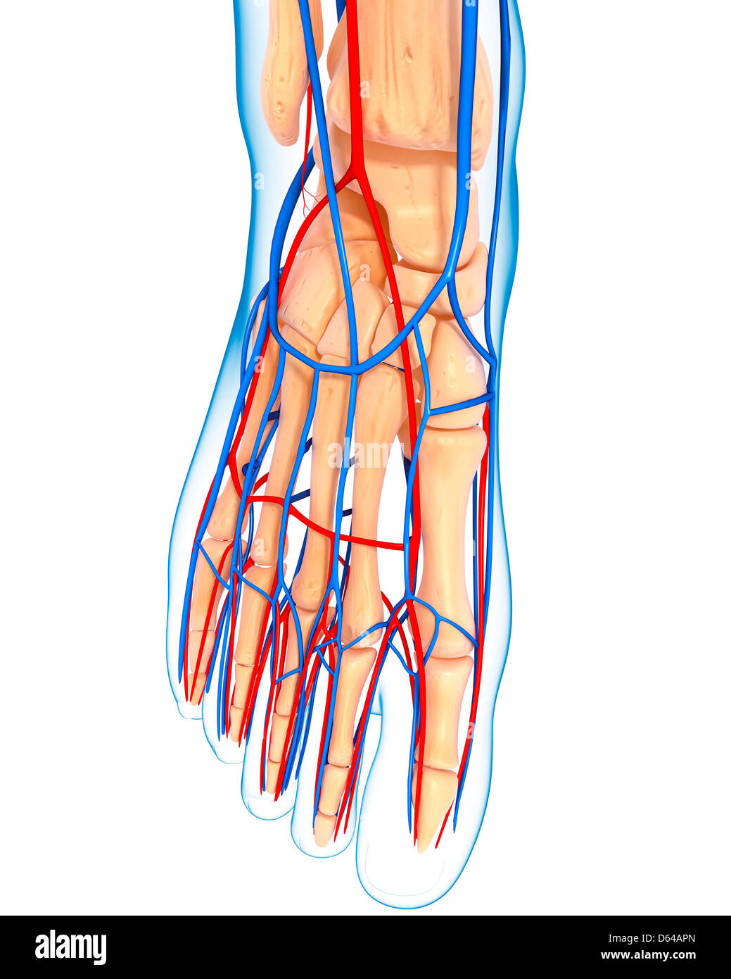 Human Foot Anatomy Stock Photos Human Foot Anatomy Stock
 Illustration Picture Of Anatomical Structures Foot Anatomy
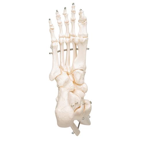 Human Foot Ankle Skeleton Wire Mounted 3b Smart Anatomy
 Ankle Joint Anatomy Overview Lateral Ligament Anatomy And

Muscles Of The Foot Summary Core Anatomy And Physiology
 Foot And Ankle Patient Education
 Foot And Ankle Anatomy Allen Tx Foot Doctor
 The Anatomical And Physiological Overview Of The Human Foot
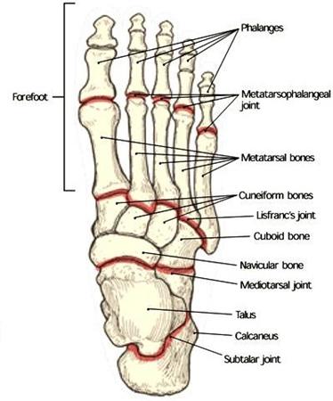 Foot Anatomy And Biomechanics Foot Ankle Orthobullets
 Foot And Ankle Anatomical Poster Size 12wx17t
Foot Anatomy Foot First Podiatry
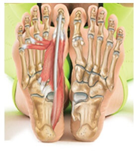 Foot And Ankle Anatomy Bones Muscles Ligaments Tendons
 Regional Anatomy Foot At Texas Woman S University Studyblue
 Illustrated Anatomy Of The Foot A The Cuneiforms Cuboid
 Foot Anatomy Detail Picture Image On Medicinenet Com
 Foot Anatomy Oceanside Ca Foot Doctor
 How To Draw Feet With Structure Foot Bone Anatomy
 Foot Anatomy Foot And Ankle Bones Ligaments Tendons And
 Foot Pain Its Anatomical Distribution Causes Of Foot Pain


Belum ada Komentar untuk "Foot Anatomy Image"
Posting Komentar