Front Neck Anatomy
The majority of these nerves control the functions of the upper extremities and allow you to feel your arms shoulder and back of your head. In the front of the neck the platysma muscle extends up from the chest goes over the collarbone and ends at the jaw.

Medially sagittal line down the midline of the neck.
Front neck anatomy. An abscessed tooth may also cause swollen lymph nodes leading to pain in the front of the neck. The suprahyoid muscles are the digastrics stylohyoid. Blood supply and vessels.
Muscles in the front of the neck are the suprahyoid and infrahyoid muscles and the anterior vertebral muscles see the images below. They are the smallest and uppermost vertebrae in the body. Sensation to the front areas of the neck comes from the roots of the spinal nerves c2 c4 and at the back of the neck from the roots of c4 c5.
Laterally anterior border of the sternocleidomastoid. The spinal column extends from the base of the skull to the pelvis. Neck anatomy nerves picture there are 8 spinal nerves that originate from the cervical spine.
It can be subdivided further into four triangles which are detailed later on in this chapter. It is also known as the pharynx. The thyroid is a butterfly shaped gland that sits low on the front of the neck.
The throat is only one part of the upper respiratory tract and digestive tract. While most of us refer to the front of the neck as the throat this is often anatomically incorrect. Your thyroid lies below your adams apple along the front of the windpipe.
It pulls down the lower face and mouth and causes wrinkles in these spots. This is a basic tutorial on the organisation of the neck describing the anatomical triangles of the neck and the different layers of fascia as well as some of the structures that pass through. In addition to nerves coming from and within the human spine the accessory nerve and vagus nerve travel down the neck.
The anterior triangle is situated at the front of the neck. This may be caused by a viral infection or in some cases a form of cancer. It connects the mouth and nasal cavity to the lower airways larynx and then trachea as well as to the esophagus food pipe.
Investing fascia covers the roof of the triangle while visceral fascia covers the floor. The neck contains seven of these known as the cervical vertebrae. Swollen lymph nodes may cause front neck pain in some people.
Superiorly inferior border of the mandible jawbone.
![]() Head And Neck Anatomy Structures Arteries And Nerves Kenhub
Head And Neck Anatomy Structures Arteries And Nerves Kenhub

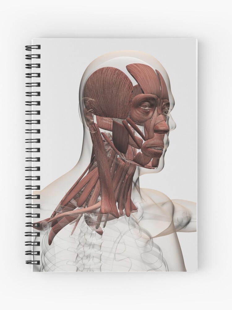
 Anatomy Of Human Face And Neck Muscles Front View Metal Print
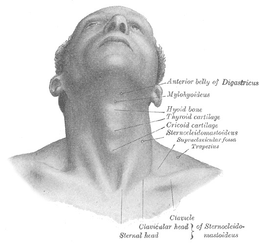 Figure Surface Anatomy And Surface Markings
 Neck Muscles Stock Illustration Illustration Of Drawing
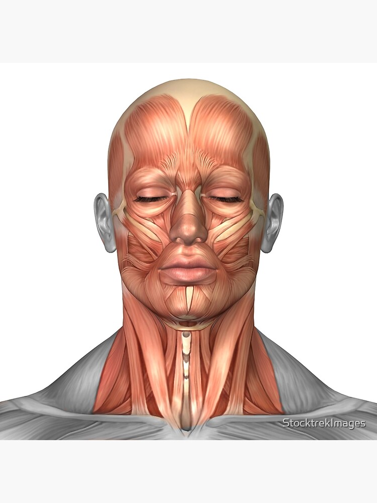
 Anatomy Of Male Facial And Neck Muscles Front View Spiral Notebook
 Front Neck Muscles Hyloids And That Stuff Ucf Youtube
 Understanding Muscles A Clue To Avoid Being A Pain In The
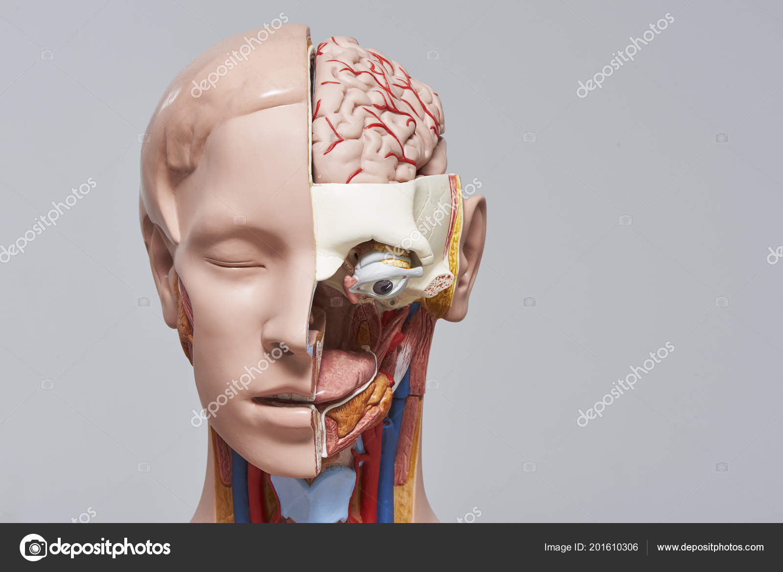 Front View Human Head Neck Model Section Showing Internal
 Female Muscular Anatomy Semi Transparent Front View Clipart
 Structure And Function Of The Cervical Spine Physiopedia
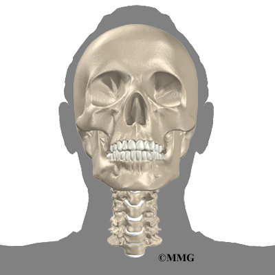
 Free Anatomy Quiz The Human Skull Quiz 1
![]() Head And Neck Anatomy Structures Arteries And Nerves Kenhub
Head And Neck Anatomy Structures Arteries And Nerves Kenhub
 Anatomy Of Human Face And Neck Muscles Front View Framed Print Wall Art By Stocktrek Images
 The Human Body Diagram Of Throat Wiring Diagram Images Gallery
 Neck Anatomy Organisation Of The Neck Part 1
 Front Neck Muscles Muscle Stretches Thyroid Health Muscle
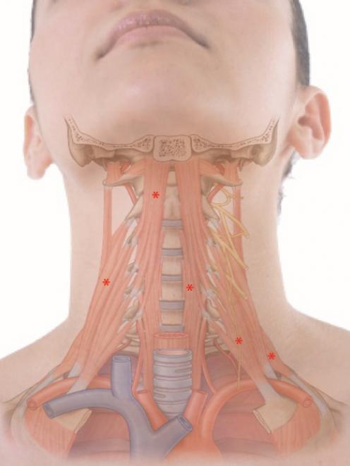
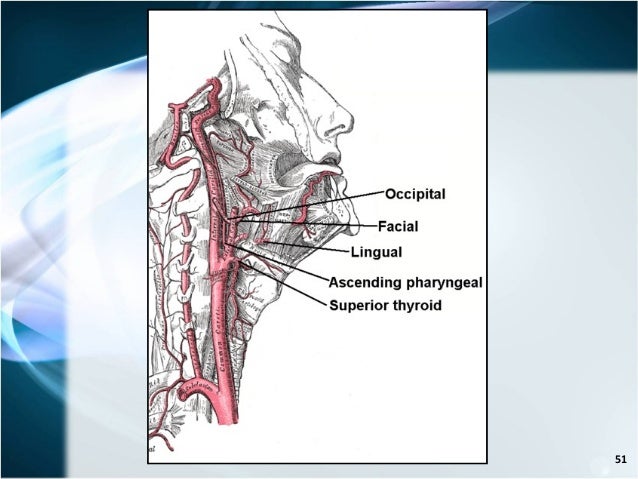
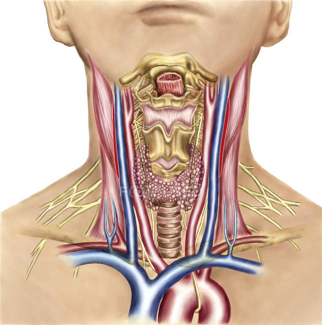 Neck Anatomy Showing Arteries Of Pharyngeal Region And
 Surgical Anatomy Of The Neck

 About Thyroid Cancer Uk Healthcare
 Throat Anatomy Anatomy Of The Larynx From The Front Top
 Anterior Shoulder Muscles Shoulder Muscle Anatomy
Belum ada Komentar untuk "Front Neck Anatomy"
Posting Komentar