Prostate Mri Anatomy
An mri study of the prostate incorporates a powerful magnet system radio waves and a computer to create very detailed images of the prostate gland and surrounding anatomy. A magnetic resonance imaging mri scanner uses strong magnetic fields to create an image or picture of the prostate and surrounding tissues.
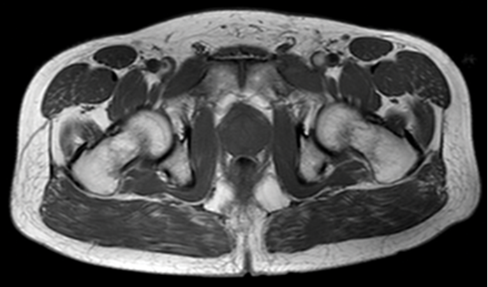
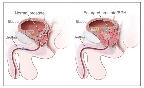
Below an example of a prostate with minimal bph 30 ml entire gland.

Prostate mri anatomy. What sequences to use. Positive digital rectal exam dre and negative trus biopsy. Define benign prostatic hypertrophy.
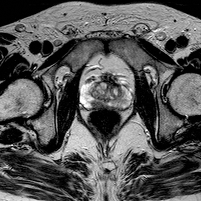
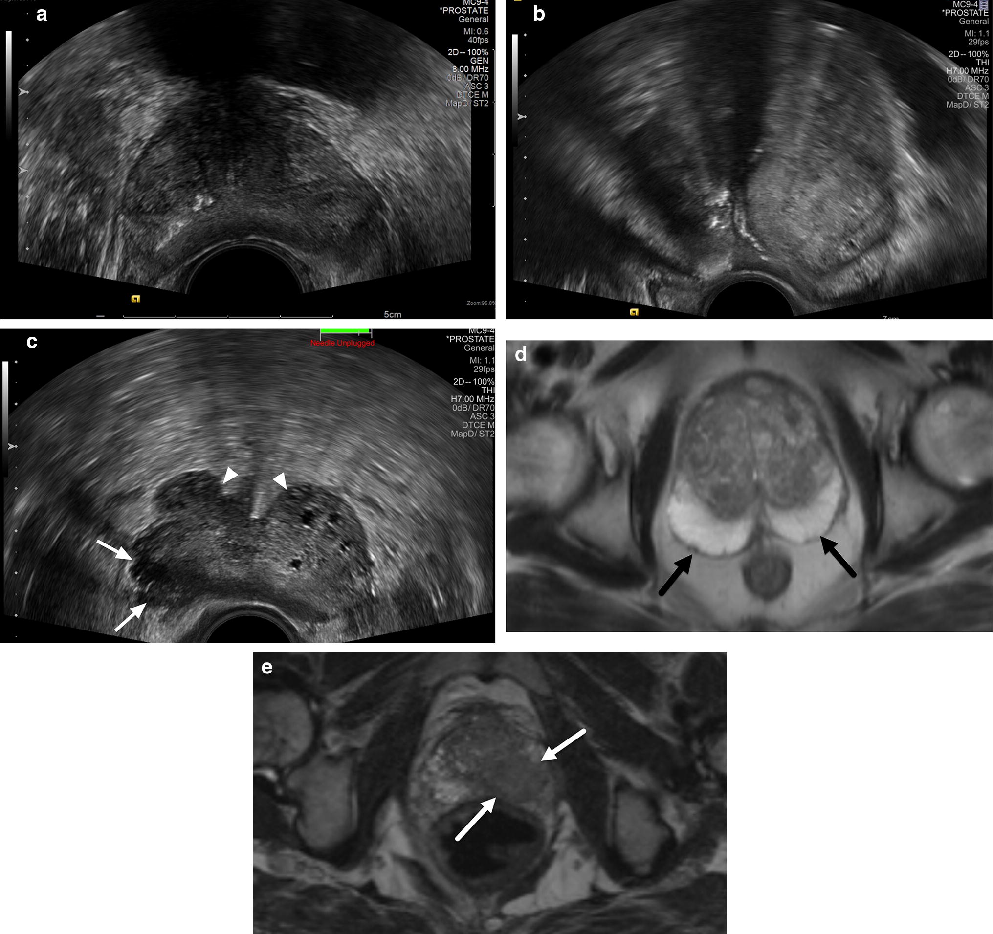
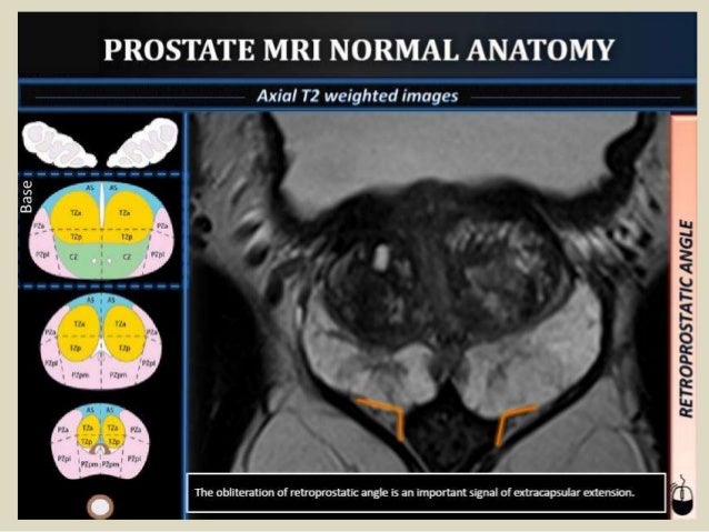
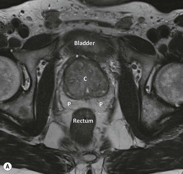
Prostate anatomy on mri. Evaluating sequential sections the peripheral central and transition zones could be differentiated. Pi rads version 2 prostate imaging reporting and data system applying pi rads scoring in the peripheral zone and transition zone.
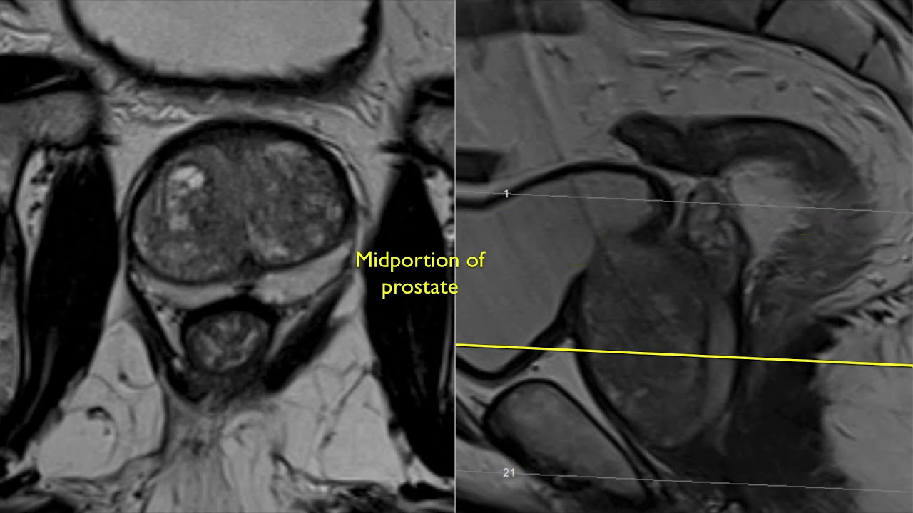
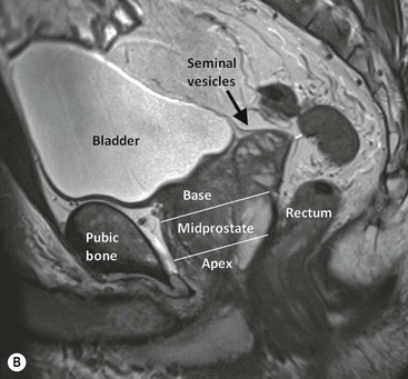
Mri of the prostate has become increasingly popular with the use of multiparametric mri and the pi rads classification. From superior to inferior the gland is commonly divided into 3 levels. Mr imaging of the prostate gland.
Multiparametric mri is a combination of t2 weighted diffusion and dynamic contrast enhanced imaging and is an accurate tool in the detection of clinically significant prostate cancer. The peripheral zone showed higher signal intensity than either the central or transition zone and was discerned in the coronal sagittal and transverse planes. Prostate mri is for men who have.
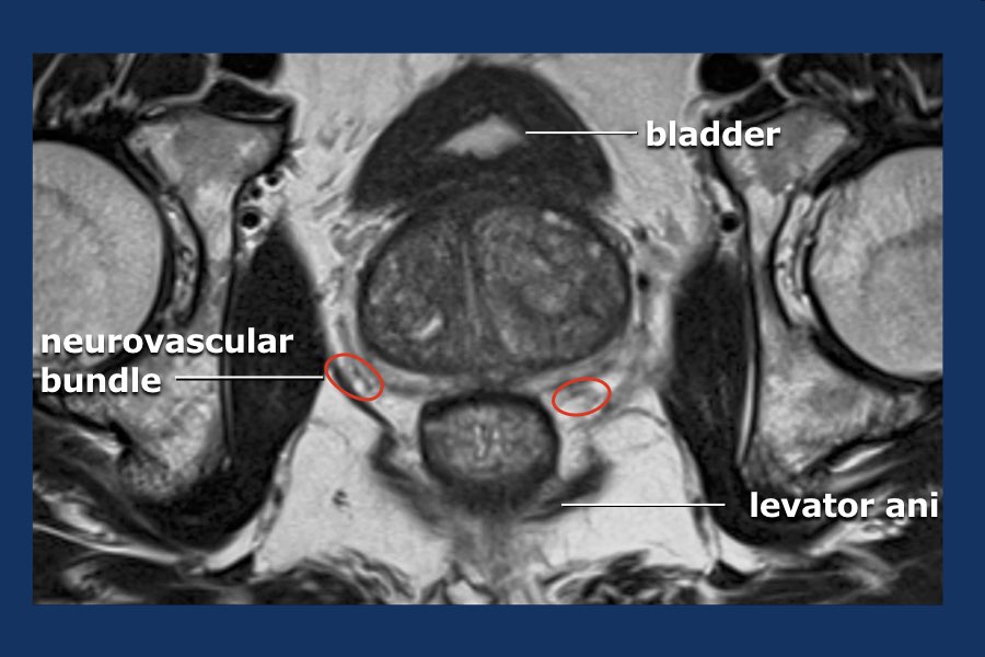
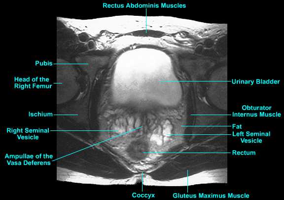
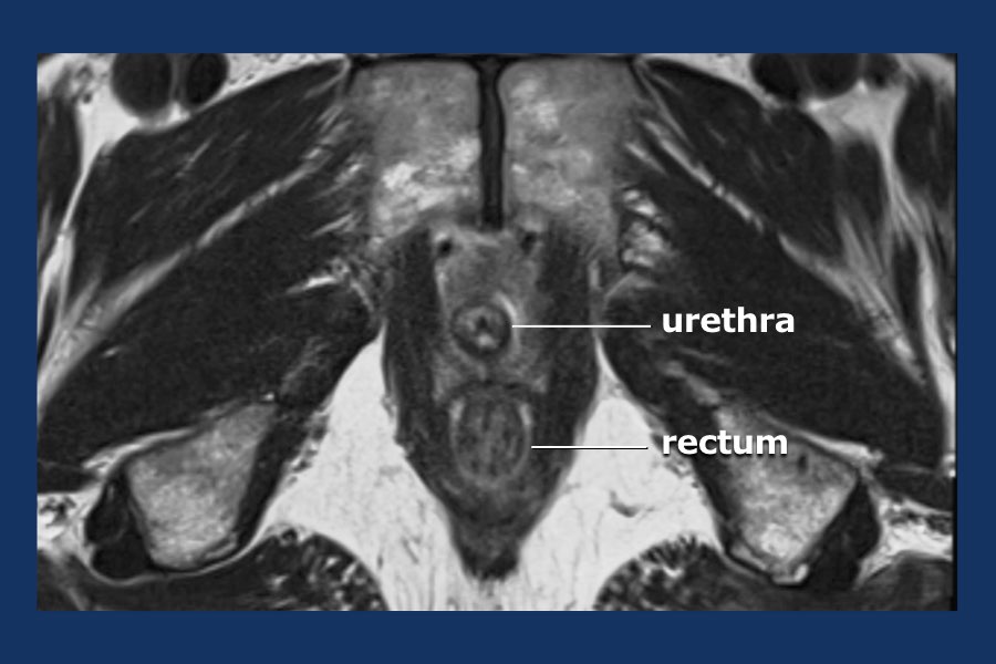
The prostate gland is a small soft structure about the size and shape of a walnut which lies deep in the pelvis between the bladder and the penis and in front of the rectum back passage. Coronal axial sagittal anatomy. The best anatomic detail is on small fov t2wi.
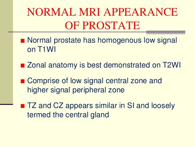
Mri imaging is helpful in differentiation the prostatic zonal anatomy best demonstrated on t2wi. Special techniques are used to improve the early detection of prostate cancer such as dwi dynamic contrast enhanced mri and mr spectroscopy. Elevated or rising prostate specific antigen psa and at least one negative transrectal ultrasound guided trus biopsy.
A prostate cancer diagnosisto provide accurate staging and guide treatment options or assess disease progression. Assessments for dce tz pz. There is no ionizing radiation x rays used to create any mri image.
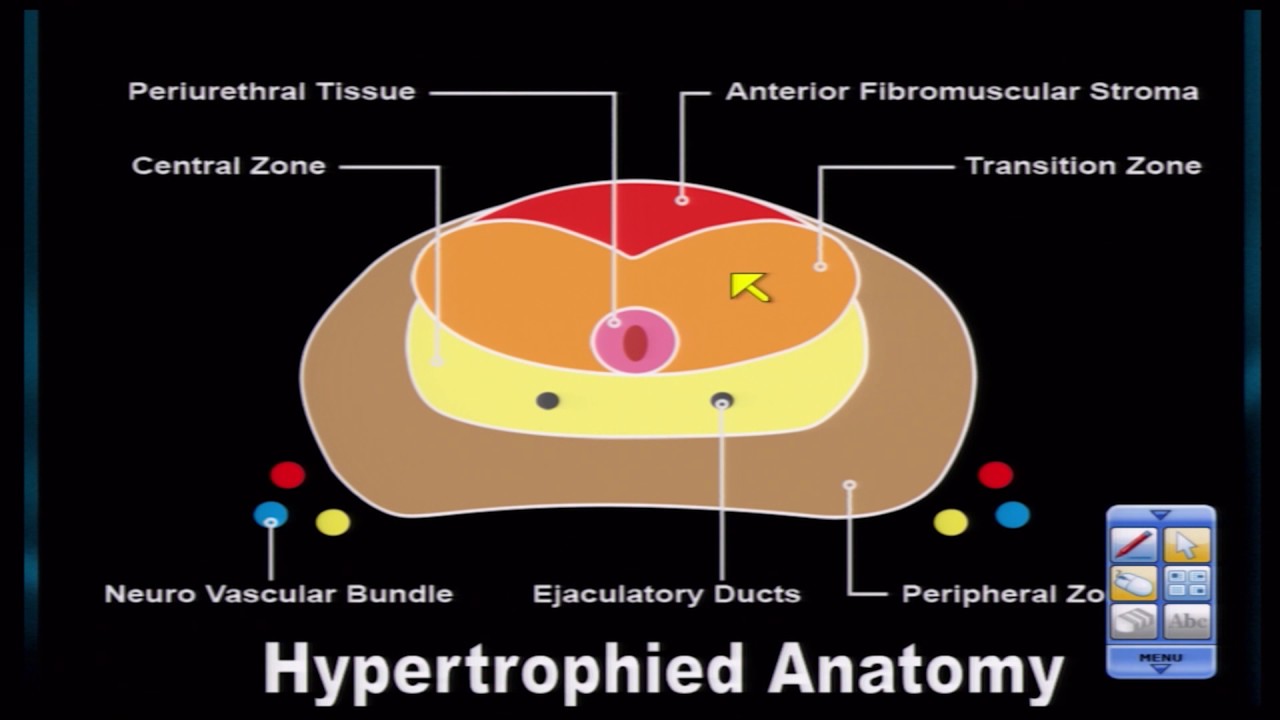
The majority of cancers arise from the peripheral zone pz.
 Diagnostics Free Full Text Anatomy And Imaging Of Rat
 The Radiology Assistant Prostate Cancer Pi Rads V2
 Normal Anatomy Of The Prostate Using Integrated Endorectal

 Imaging In Prostate Cancer
 Use Of Mri In The Evaluation Of Prostate Cancer Part 1
 Use Of Mri In The Evaluation Of Prostate Cancer Part 1
 Prostate Imaging Borg Ide Imaging
 Prostate Cancer Detection And Diagnosis Role Of Ultrasound
 Artificial Intelligence For Mri Siemens Healthineers Global
 Prostate Anatomy Mri
 Normal Prostate Anatomy On Magnetic Resonance Imaging T1 And
 The Radiology Assistant Prostate Cancer Pi Rads V2
 Mri Prostate
 Presentation1 Mri Imaging Of The Prostate

Multiparametric 3t Mr Imaging Of The Prostate Acquisition
Gallery Magnetic Resonance Imaging Mri Image 997
 Prostate Mri Case Review A Continued Look At Anatomy 30

 The Male Pelvis Mr Anatomy Atlas Of The Prostate Bladder
 The Hip Anatomy On 3t Mr And 3d Pictures
 Prostate Radiology Key
 Multiparametric Mr Imaging In Diagnosis Of Chronic
 Normal Prostate Mri Radiology Case Radiopaedia Org

 Prostate Radiology Key
 Prostate Mr Stages Of Prostate Cancer Ustoo
 Mri Pelvis Anatomy Free Male Pelvis Axial Anatomy

Belum ada Komentar untuk "Prostate Mri Anatomy"
Posting Komentar