Shin Anatomy
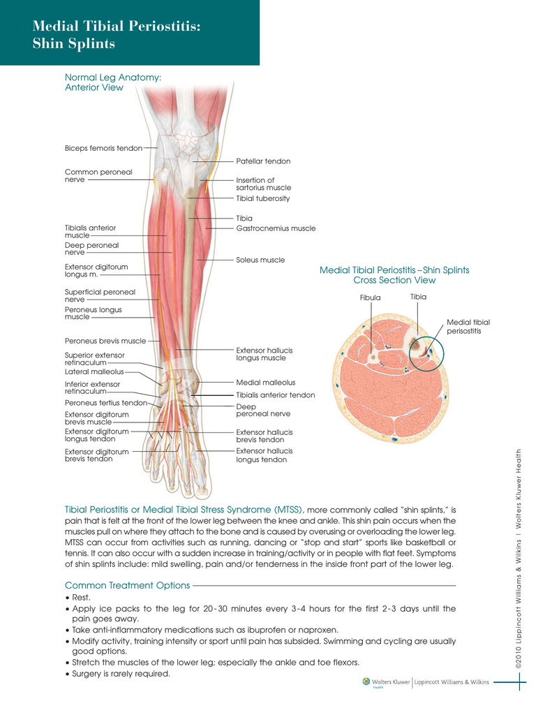
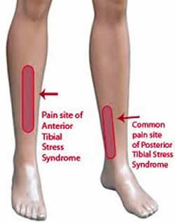
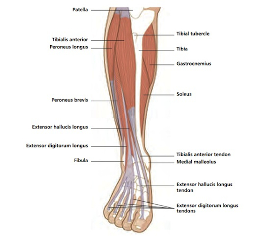
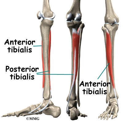
Pain typically occurs along the inner border of the tibia where muscles attach to the bone. The tibialis anterior is a muscle in humans that originates in the upper two thirds of the lateral surface of the tibia and inserts into the medial cuneiform and first metatarsal bones of the foot.
 Anatomy Of Shin Splints Treatment And Prevention
It acts to dorsiflex and invert the foot.
Shin anatomy. Anatomy of shin splints. The most common symptom of shin splints is pain. Treatment and prevention description.
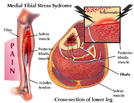
In general shin splints develop when the muscle and bone tissue periosteum. The lateral compartment is along the outside of the lower leg. Shin splints medial tibial stress syndrome is an inflammation of the muscles tendons and bone tissue around your tibia.
The muscles of the calf also work subtly to stabilize the ankle joint and foot and to maintain the bodys balance. The tibialis anterior overlaps the anterior tibial vessels and deep peroneal nerve in the upper part of the leg. Shin splint anatomy and treatment mark charrette dc april 18 2019 shin splints or medial tibial stress syndrome has become a household word as more people pursue exercise for recreation and better health.
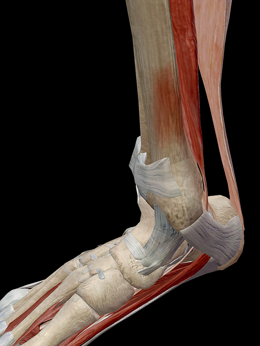
The dermis beneath the epidermis contains tough connective tissue hair follicles and sweat. The deep posterior compartment. The muscles of the lower leg the anterior compartment in the front of the shin holds the tibialis anterior.
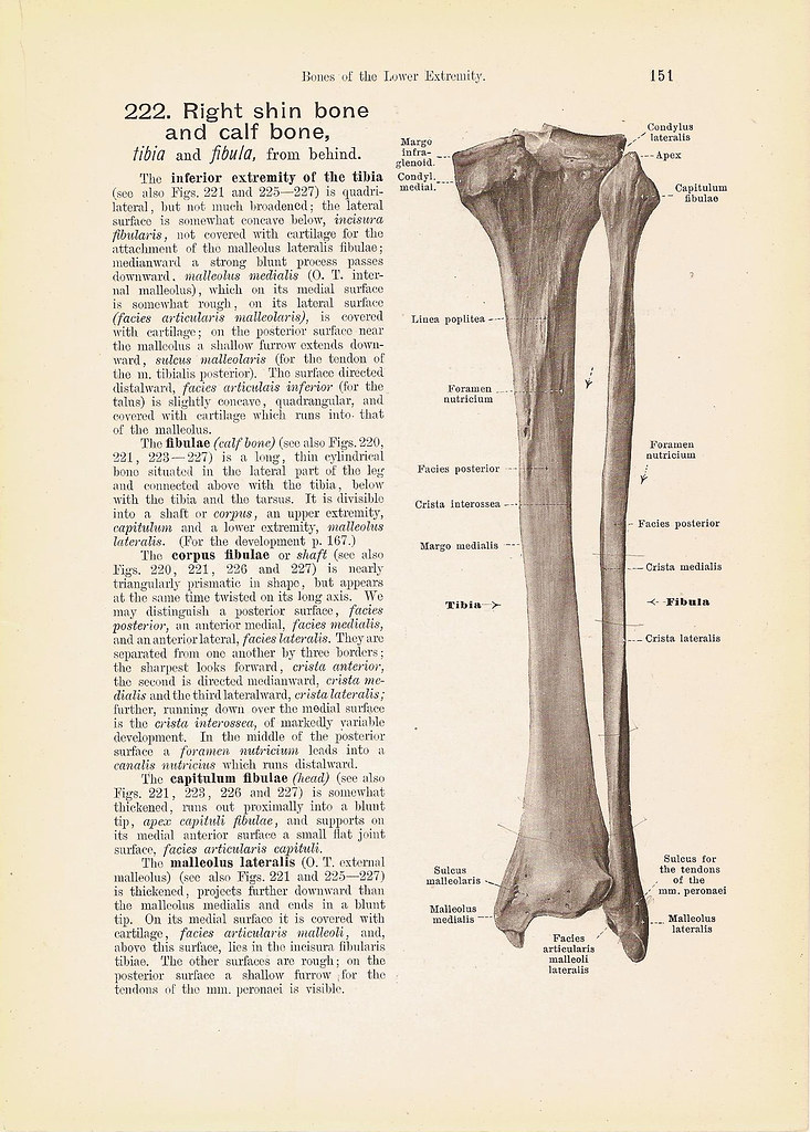
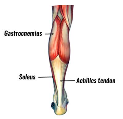
The tibia also called the shinbone is located near the midline of the leg and is the thicker and stronger of the two bones. The posterior compartment holds the large muscles that we know as the calf muscles. It is situated on the lateral side of the tibia.
The fibula also called the calf bone is significantly smaller and is. Shin muscles such as the tibialis anterior and extensor digitorum longus dorsiflex the foot and extend the toes. The tibia ˈ t ɪ b i ə plural tibiae ˈ t ɪ b i i or tibias also known as the shinbone or shankbone is the larger stronger and anterior frontal of the two bones in the leg below the knee in vertebrates the other being the fibula behind and to the outside of the tibia and it connects the knee with the ankle bones.
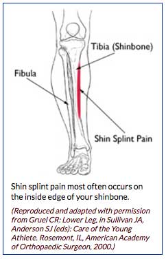
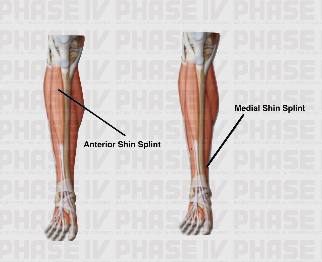
Cookery chiefly brit a cut of beef the lower foreleg. Shin splint pain most often occurs on the inside edge of your tibia shinbone. The epidermis the outermost layer of skin provides a waterproof barrier and creates our skin tone.
Shin splints medial tibial stress syndrome is an inflammation of the muscles. This muscle is mostly located near the shin. Anatomy the front edge of the tibia.
It is thick and fleshy above tendinous below. Anatomy the front part of the lower leg.
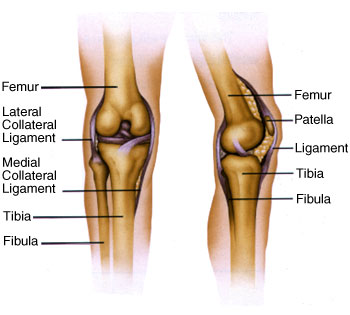 Knee Anatomy

 What Is Shin Splints And Are You Suffering From It Know
 Shin Splints
 Symptom Of The Month March
 Thigh Calf And Shin Zamst
 Hold On To Your Tibias The Anatomy And Causes Of Shin Splints
 Shin Splints


 Shin Splints Orthogate
 Right Shin Calf Tibia And Fibula Engraving Anatomy Book
 Human Leg Wikipedia

 Calf Strain Torn Calf Muscle Treatment Rehabilitation
 Tibias On Fire Shin Splints
 Pin On Anatomy
 Shin Bone Art Page 4 Of 6 Fine Art America
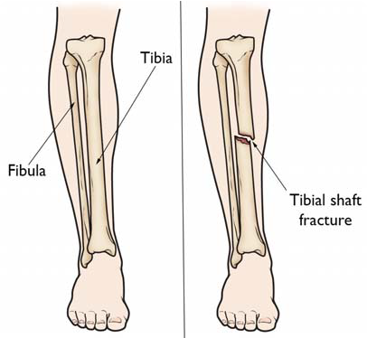
Shinbone Fracture Boston Medical Center
Belum ada Komentar untuk "Shin Anatomy"
Posting Komentar