Microscope Anatomy
For resolving the details of objects which otherwise cannot be achieved by naked eye a microscope is used. The most common type of modern microscope is called a compound microscope.

This type of microscope has become so advanced that some are capable of magnifying up to 1000 times.

Microscope anatomy. Moves the stage up down in order to bring the image into view. Stages are often equipped with a mechanical device that holds the specimen slide in place and can smoothly translate the slide back and forth as well as from side to side. The study of the microscopic structure of the tissues and cells.
This test will help the student to identify the different parts and function of the microscope. Kinds of microscopic anatomy are cytology and histology. They have two systems of lenses one is the eyepiece and the other is comprised of one or more objective lenses.
Holds the objectives and can be rotated to change the magnification. It works like a lazy susan. It contains one lens which has the magnification power of 10x.
Students can view and utilize these tutorials using a web browser without the addition of plug in software. Microscopes are instruments designed to produce magnified visual or photographic images of small objects. The microscope must accomplish three tasks.
Use only when scope is on low power. The objective lenses are mounted in this part of the microscope. A list of the fundamental parts which make up the anatomy of a microscope.
This is a device which amplifies and magnifies the view upon a subject. Microscope anatomy interactive java tutorials we have constructed a variety of interactive java driven microscopy tutorials to help explain some of the more difficult concepts in optical microscopy. The user looks through this.
Anatomy of the microscope. All microscopes are designed to include a stage where the specimen usually mounted onto a glass slide is placed for observation. A microscope is an instrument widely to magnify and resolve the image of an object that is otherwise invisible to naked eye.
Produce a magnified image of the specimen separate the details in the image and render the details visible to the human eye or camera.
 200 Prepared Microscope Slides Specimen Set Plant Animal Human Anatomy Cells
Anatomy Of A Microscope Ppt Download
 Usd 1298 57 式式 显微镜 显微镜 解剖 解剖 解剖 立体 显微镜
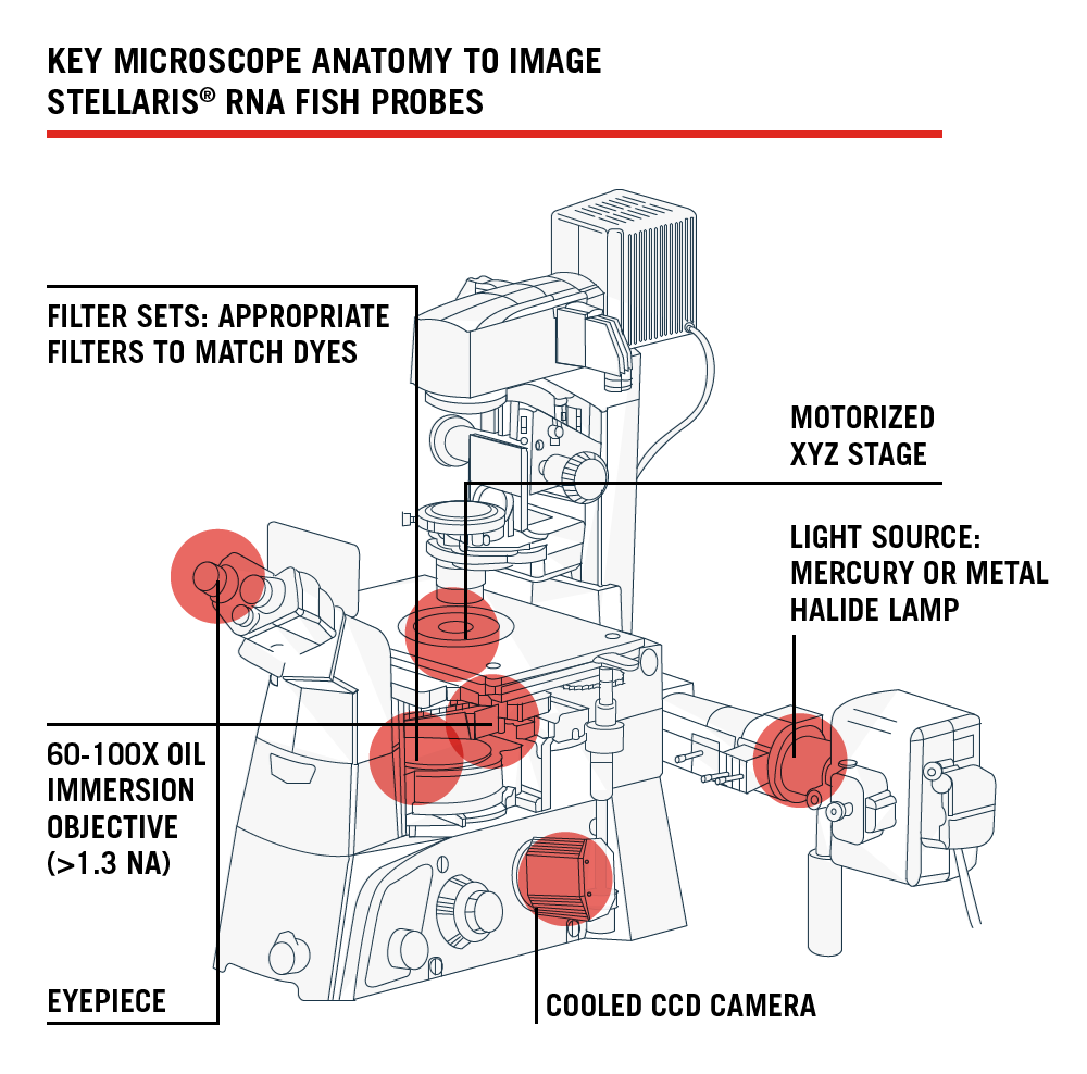
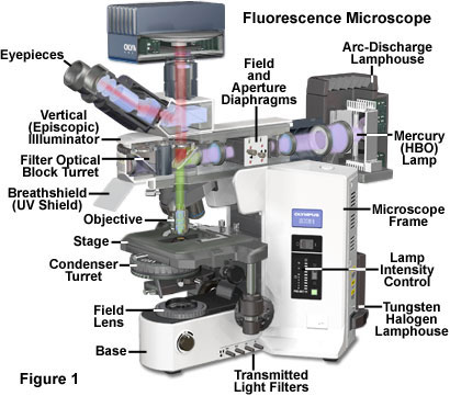 Fluorescence Microscopy Anatomy Of The Fluorescence
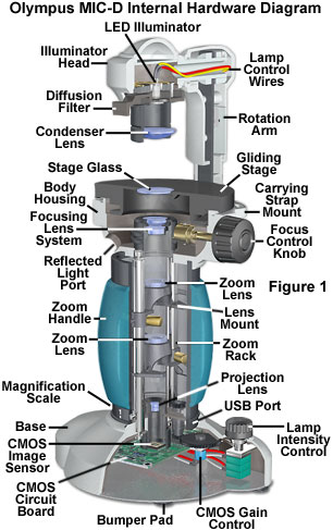 Anatomy Of The Mic D Digital Microscope Overview Olympus
 Chapter 3 Solutions Human Anatomy Physiology Laboratory
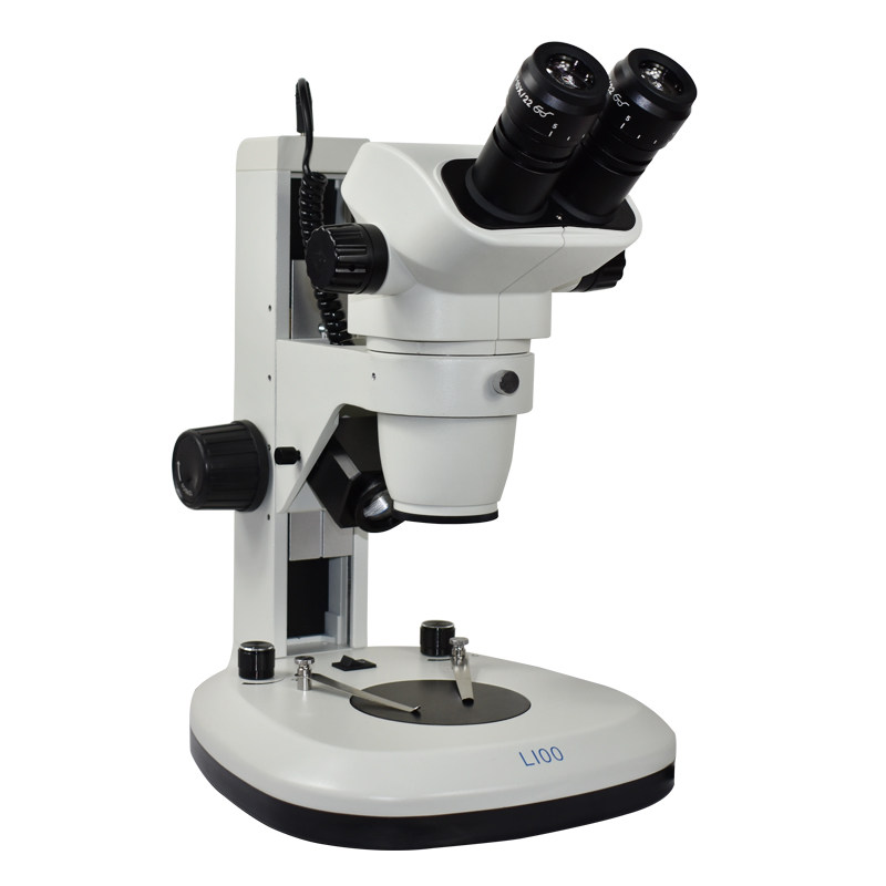
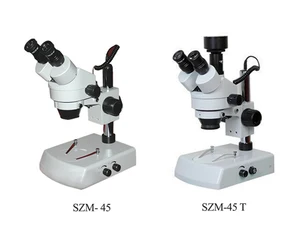 Stereo Microscope For Medical Anatomy Analysis
 Microscope Anatomy Diagram Quizlet
 2019 Ba 008t Hd Biological Microscope Upper And Lower Light Source Jewelry Identification Specimen Anatomical Body Magnifying Glass From Baisidatools
 Microscopy Anatomy Microscope Dissection Biology
 Us 109 73 15 Off 20x 40x Zoom Dissecting Stereology Stereo Microscope With Top And Bottom Illumination For Maintenance Anatomy Jewelry Appraisal In
 Revive An Old Microscope Proper Cleaning New Light Source
 Glossary Greatscopes
What Is Fluorescence Microscopy Quora
 Components Of Meiji Microscope Anatomy Physiology 1086

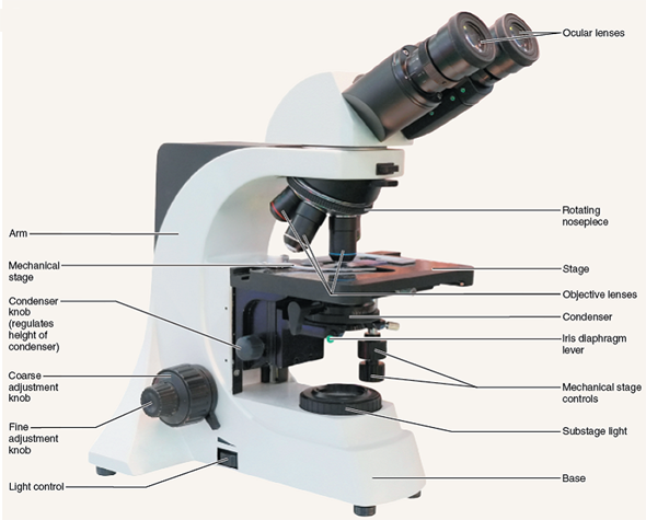
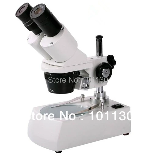
 Compound Microscope Parts
 Amazon Com Microscope Black White Medical Art Print
 Olympus Orbeye Video Microscope To Appear On Abc S Grey S
 Parts Of A Compound Microscope With Diagram And Functions
 Lab Equipment Anatomy And Terminology Of Microscopes From
 This Is A Quiz Called Microscope Labeling Game And Was
 Revive An Old Microscope Proper Cleaning New Light Source
 Anatomy Of A Microscope In Uf Microbiology On Vimeo
 The B H Microscope Buying Guide B H Explora
Belum ada Komentar untuk "Microscope Anatomy"
Posting Komentar