Ramus Anatomy
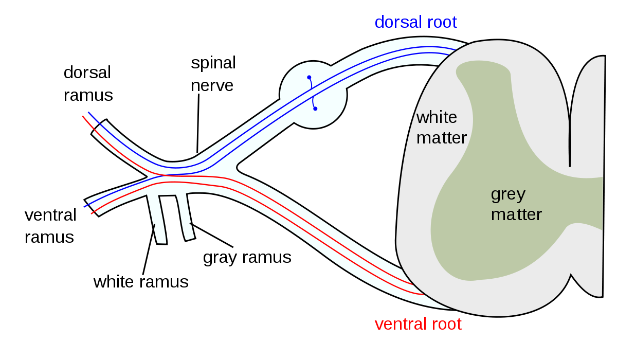
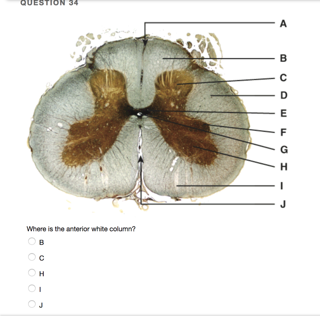
For example the ramus acetabularis arteriae circumflexae femoris medialis is the branch of an artery that goes to the socket of the hip joint. The dorsal ramus latin for branch plural rami is the dorsal branch of a spinal nerve that forms from the dorsal root of the nerve after it emerges from the spinal cord.
 Male Vs Female Pelvis Differences Anatomy Of Skeleton
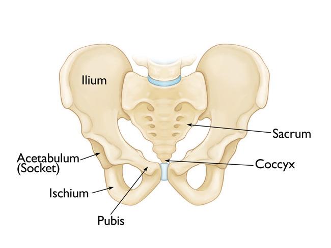
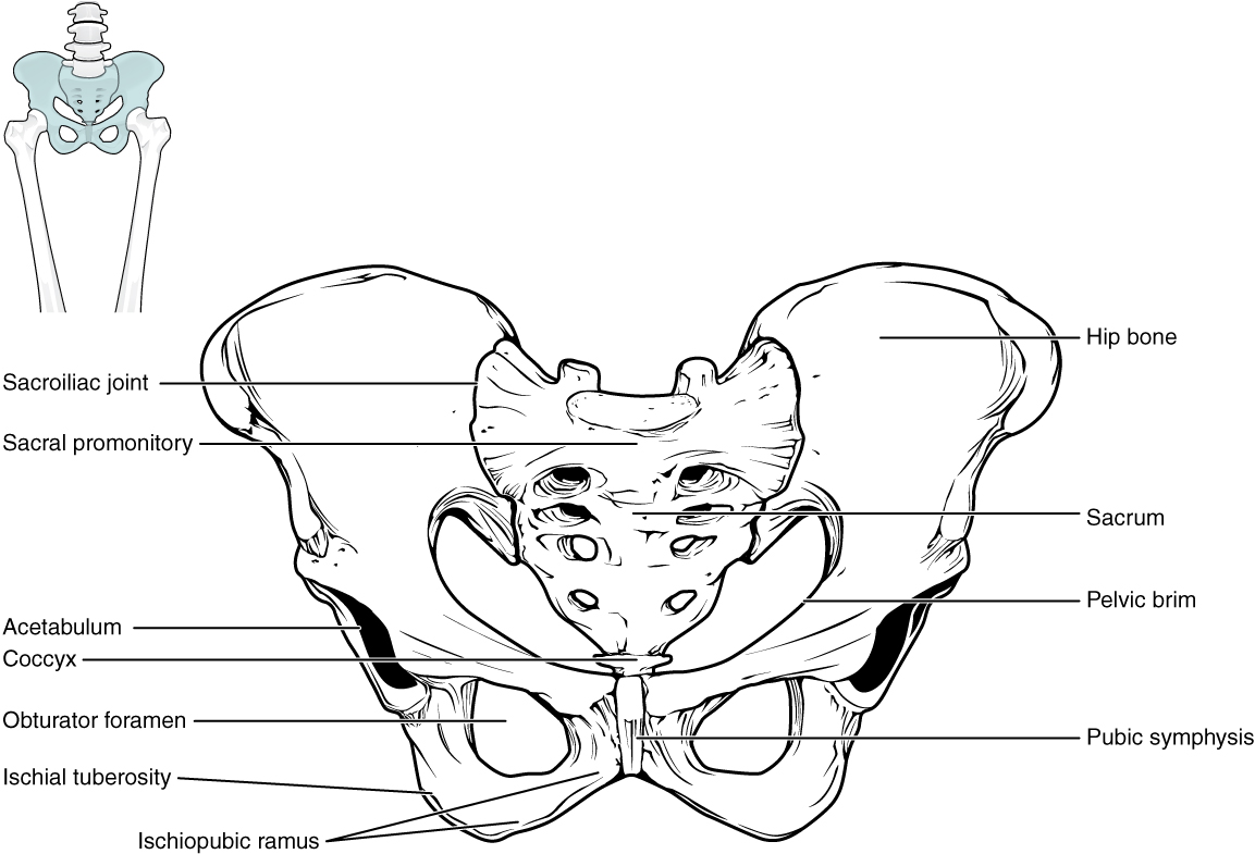
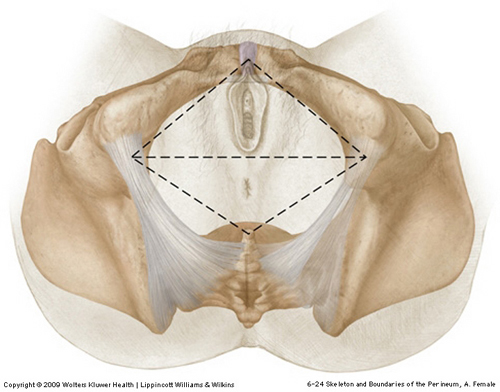
Rami is a part of the pubic bone which forms a portion of the obturator foramen.

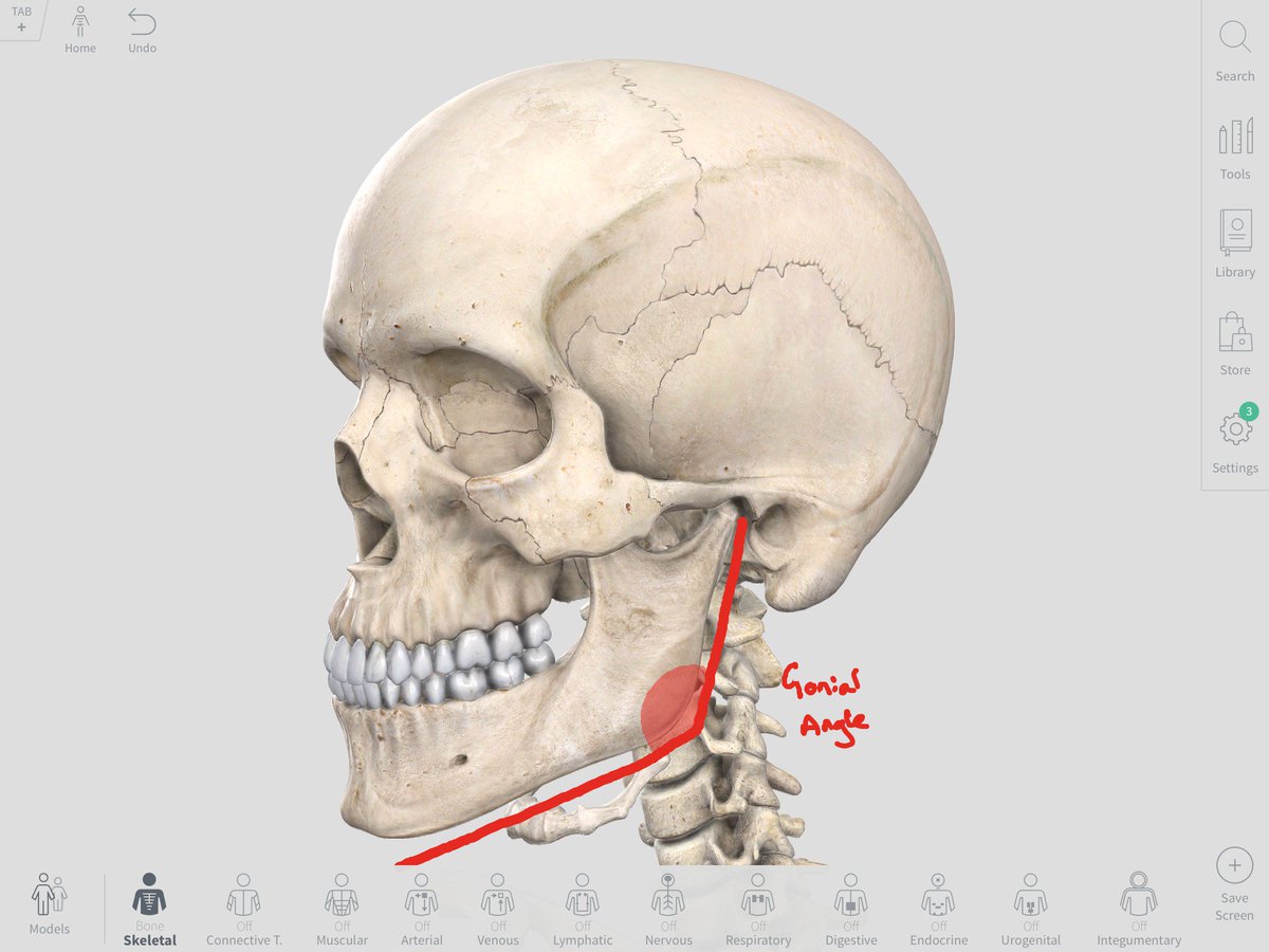
Ramus anatomy. In anatomy a branch such as a branch of a blood vessel or nerve. The joints by means of which the lower jaw is. The superior pubic ramus consists of two parts.
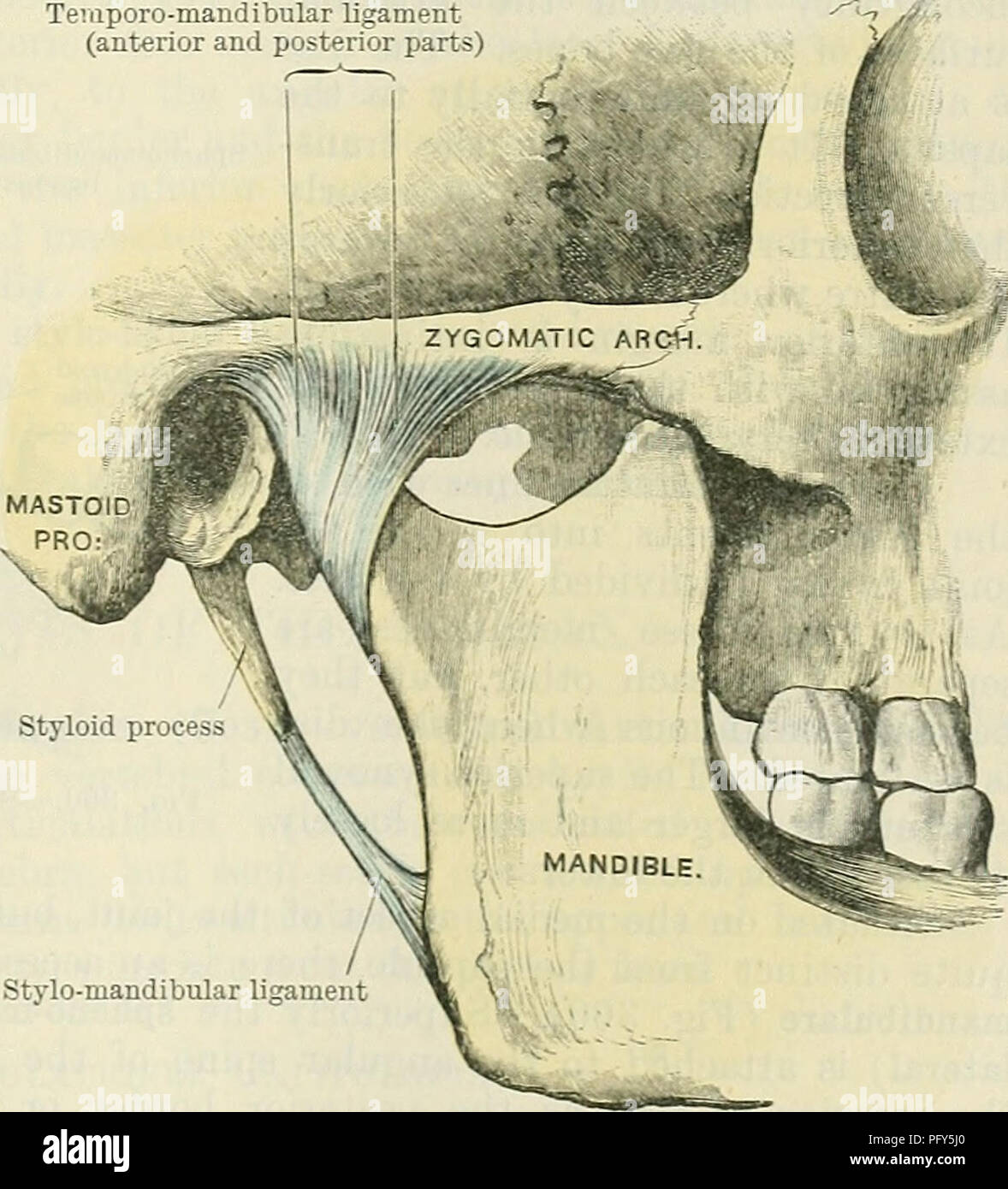
The superior pubic ramus connects with the ilium along with ischium at its base which is located toward the acetabulum and protrudes posterolaterally from the body. Jaw structure in jaw two vertical portions rami form movable hinge joints on either side of the head articulating with the glenoid cavity of the temporal bone of the skull. A branch as of a nerve or blood vessel or a projecting part as of a rotifer or crustacean.
The incidence is some where between 10 30. The dorsal ramus carries information that supplies muscles and sensation to the human back. It is present in 20 range 15 30 2 3 of the population.
The medial part consists of two surfaces anterior posterior and three borders upper medial lateral. A medial part the body of the pubis and a lateral part. The spinal nerve is formed from the dorsal and ventral rami.
It can have a course similar to the obtuse marginal branches of the le. A bony process extending like a branch from a larger bone especially the ascending part of the lower jaw that makes a joint at the temple. It extends from the body to the median plane where it articulates with its fellow of the opposite side.
The obturator foramen along with the ilium and other fused bones forms part of either side of the pelvis. Any of the primary divisions of a nerve or blood vessel. The ramus intermedius is a variant coronary artery resulting from trifurcation of the left main coronary artery 1.
Normally the main coronary blood vessel has two branches the left anterior descending artery and left circumflex but some people have a third branch termed intermediate artery or ramus coronary artery see picture. The sharp superior border of triangular surface creates part of the linea terminalis of the pelvic bone as well as the pelvic inlet and is called the pectineal line aka. The superior pubic ramus projects posteriorly and laterally from the body and joins the ilium and ischium.
The superior pubic ramus pl. A part of an irregularly shaped bone that is thicker than a process and forms an angle with the main body especially the ascending part of the lower jaw that makes a joint at the temple. Any of the primary divisions of a cerebral sulcus.
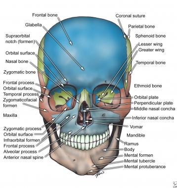
The lower jaw at the side are called rami branches.
Ischial Prolotherapy Journal Of Prolotherapy
 Cunningham S Text Book Of Anatomy Anatomy Akticulatiox Of
 Facial Bone Anatomy Overview Mandible Maxilla
 Pelvic Fractures Orthoinfo Aaos
 Ventral Ramus Of Spinal Nerve Wikipedia
 Cervical Vertebra Lateral View Human Anatomy Handout
 File An Atlas Of Human Anatomy For Students And Physicians
 Genital Branch Of Genitofemoral Nerve Wikipedia
 Anatomy Of The Occipital And Suboccipital Regions 1 Head
Imaging Mandibular Fractures Wikiradiography
 Solved Question 30 Where Is The Posterior Ramus None Of
 Anatomy Test 3 Spinal Cord And Nerves Diagram Quizlet
 Dr Stephen Maclean On Twitter My Fun Anatomy Fact For
 Sylvian Fissure Radiology Reference Article Radiopaedia Org
 Spinal Nerve Anatomy Britannica
 Inferior Frontal Gyrus Wikipedia
:watermark(/images/watermark_5000_10percent.png,0,0,0):watermark(/images/logo_url.png,-10,-10,0):format(jpeg)/images/atlas_overview_image/441/1Mvvs4WAYFJHq6jT2EZCkg_spinal-cord-in-situ_english.jpg) Sympathetic Nervous System Definition Anatomy Function
Sympathetic Nervous System Definition Anatomy Function
 The Anterior Divisions Human Anatomy
Pelvis And Perineum Gray S Anatomy Medicine Mbchb Studocu

 8 3 The Pelvic Girdle And Pelvis Anatomy And Physiology
 Anatomy Of A Thoracic Spinal Nerve With The Intercostal
 Cervical Plexus And Greater Occipital Nerve Blocks


Belum ada Komentar untuk "Ramus Anatomy"
Posting Komentar